Publication
Metrics
AI Quick Summary
The proposed architecture outperforms existing methods by achieving the highest mean intersection-over-union score, facilitating more efficient evaluation of surgical excision completeness.
Paper Preview
Abstract
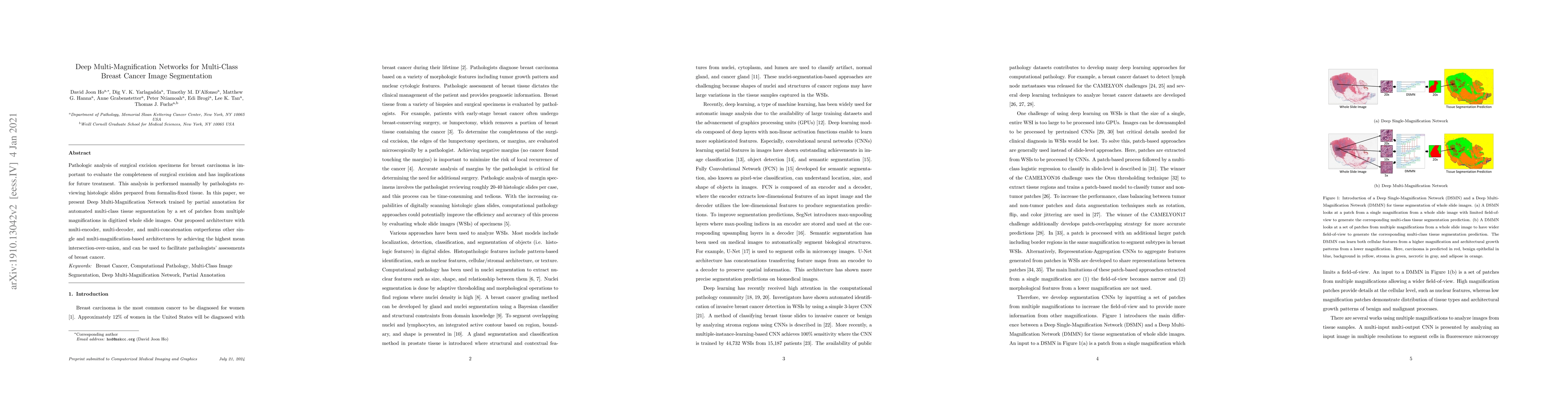
Pathologic analysis of surgical excision specimens for breast carcinoma is important to evaluate the completeness of surgical excision and has implications for future treatment. This analysis is performed manually by pathologists reviewing histologic slides prepared from formalin-fixed tissue. In this paper, we present Deep Multi-Magnification Network trained by partial annotation for automated multi-class tissue segmentation by a set of patches from multiple magnifications in digitized whole slide images. Our proposed architecture with multi-encoder, multi-decoder, and multi-concatenation outperforms other single and multi-magnification-based architectures by achieving the highest mean intersection-over-union, and can be used to facilitate pathologists' assessments of breast cancer.
AI Key Findings
Get AI-generated insights about this paper's methodology, results, significance, and more — seven facets brought into focus.
Impact
Paper Details
PDF Preview
Key Terms
Citation Network
Current paper (gray), citations (green), references (blue)
Display is limited for performance on very large graphs.
Discussion 0