Deep Superpixel Generation and Clustering for Weakly Supervised Segmentation of Brain Tumors in MR Images
Publication
Metrics
AI Quick Summary
This paper proposes a weakly supervised method for brain tumor segmentation using deep superpixel generation and clustering, which leverages binary image-level classification labels instead of detailed ground truth annotations. The method achieved a mean Dice coefficient of 0.691 and a mean 95% Hausdorff distance of 18.1, outperforming existing superpixel-based methods.
Paper Preview
Abstract
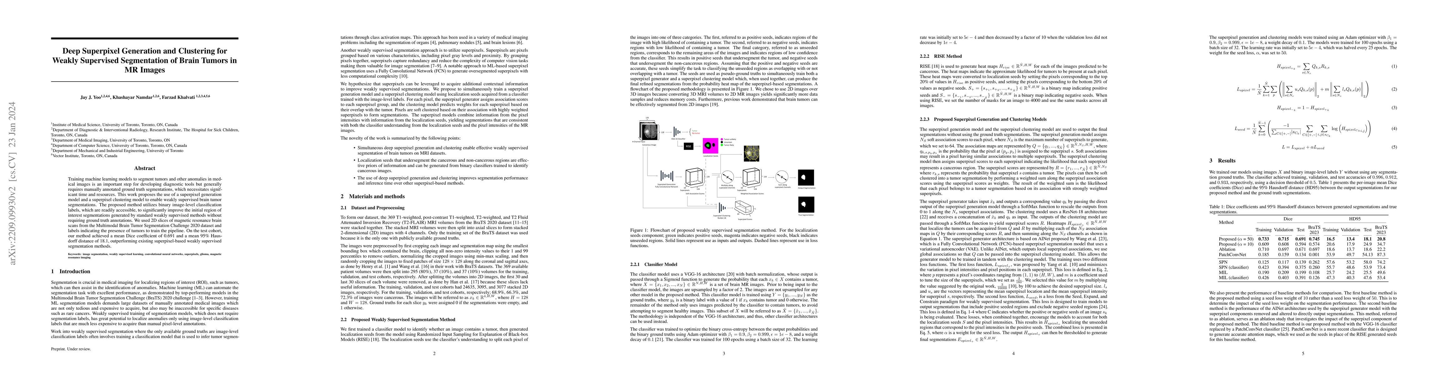
Training machine learning models to segment tumors and other anomalies in medical images is an important step for developing diagnostic tools but generally requires manually annotated ground truth segmentations, which necessitates significant time and resources. This work proposes the use of a superpixel generation model and a superpixel clustering model to enable weakly supervised brain tumor segmentations. The proposed method utilizes binary image-level classification labels, which are readily accessible, to significantly improve the initial region of interest segmentations generated by standard weakly supervised methods without requiring ground truth annotations. We used 2D slices of magnetic resonance brain scans from the Multimodal Brain Tumor Segmentation Challenge 2020 dataset and labels indicating the presence of tumors to train the pipeline. On the test cohort, our method achieved a mean Dice coefficient of 0.691 and a mean 95% Hausdorff distance of 18.1, outperforming existing superpixel-based weakly supervised segmentation methods.
AI Key Findings
Get AI-generated insights about this paper's methodology, results, significance, and more — seven facets brought into focus.
Impact
Paper Details
Authors
PDF Preview
Key Terms
Citation Network
Current paper (gray), citations (green), references (blue)
Display is limited for performance on very large graphs.
Discussion 0