DeepSPV is a novel deep learning pipeline that estimates 3D spleen volume from 2D ultrasound images, achieving high accuracy and interpretability. It outperforms human experts in spleen volume estimation and can provide confidence intervals, aiding clinicians in diagnosing splenomegaly, especially in regions lacking advanced imaging tools.
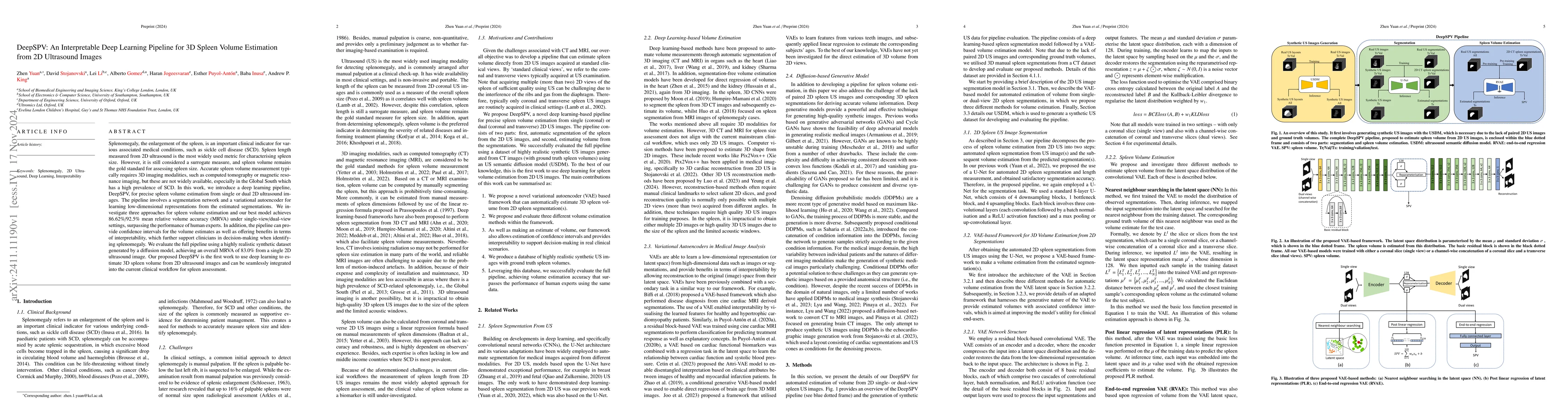
Splenomegaly, the enlargement of the spleen, is an important clinical indicator for various associated medical conditions, such as sickle cell disease (SCD). Spleen length measured from 2D ultrasound is the most widely used metric for characterising spleen size. However, it is still considered a surrogate measure, and spleen volume remains the gold standard for assessing spleen size. Accurate spleen volume measurement typically requires 3D imaging modalities, such as computed tomography or magnetic resonance imaging, but these are not widely available, especially in the Global South which has a high prevalence of SCD. In this work, we introduce a deep learning pipeline, DeepSPV, for precise spleen volume estimation from single or dual 2D ultrasound images. The pipeline involves a segmentation network and a variational autoencoder for learning low-dimensional representations from the estimated segmentations. We investigate three approaches for spleen volume estimation and our best model achieves 86.62%/92.5% mean relative volume accuracy (MRVA) under single-view/dual-view settings, surpassing the performance of human experts. In addition, the pipeline can provide confidence intervals for the volume estimates as well as offering benefits in terms of interpretability, which further support clinicians in decision-making when identifying splenomegaly. We evaluate the full pipeline using a highly realistic synthetic dataset generated by a diffusion model, achieving an overall MRVA of 83.0% from a single 2D ultrasound image. Our proposed DeepSPV is the first work to use deep learning to estimate 3D spleen volume from 2D ultrasound images and can be seamlessly integrated into the current clinical workflow for spleen assessment.
Get AI-generated insights about this paper's methodology, results, and significance.
Current paper (gray), citations (green), references (blue)
Esther Puyol-Anton, Andrew P. King, Zhen Yuan et al.
Behdad Dashtbozorg, Tiziano Natali, Liza M. Kurucz et al.
No citations found for this paper.
Title | Authors | Year | Actions |
---|---|---|---|
Echo from noise: synthetic ultrasound image generation using diffusion models for real image segmentation
|
D. Stojanovski, Uxio Hermida, P. Lamata, A. Beqiri, A. Gómez
|
2023
|
View Paper |
RecON: Online learning for sensorless freehand 3D ultrasound reconstruction
|
Mingyuan Luo, Xin Yang, Hongzhang Wang, Haoran Dou, Xindi Hu, Yuhao Huang, N. Ravikumar, Songcheng Xu, Yuanji Zhang, Yi Xiong, Wufeng Xue, Alejandro F Frangi, Dong Ni, Litao Sun
|
2023
|
View Paper |
Variational autoencoder-based estimation of chronological age and changes in morphological features of teeth
|
Su-Bin Joo, Won Jung, Seung Eel Oh
|
2023
|
View Paper |
Test–retest reliability of splenic volume assessment by ultrasonography
|
Pontus K Holmström, Frank Pernett, E. Schagatay
|
2022
|
View Paper |
Conversion Between CT and MRI Images Using Diffusion and Score-Matching Models
|
Qing Lyu, Ge Wang
|
2022
|
View Paper |
Semantic Image Synthesis via Diffusion Models
|
Weilun Wang, Jianmin Bao, Wen-gang Zhou, Dongdong Chen, Dong Chen, Lu Yuan, Houqiang Li
|
2022
|
View Paper |
Attri-VAE: Attribute-based interpretable representations of medical images with variational autoencoders
|
Irem Cetin, Maialen Stephens, O. Camara, M. Ballester
|
2022
|
View Paper |
Image Super-Resolution via Iterative Refinement
|
Chitwan Saharia, Jonathan Ho, William Chan, Tim Salimans, David J. Fleet, Mohammad Norouzi
|
2021
|
View Paper |
Cascaded Regression Neural Nets for Kidney Localization and Segmentation-free Volume Estimation
|
Mohammad Arafat Hussain, G. Hamarneh, Rafeef Garbi
|
2021
|
View Paper |
Generating Synthetic Labeled Data From Existing Anatomical Models: An Example With Echocardiography Segmentation
|
A. Gilbert, M. Marciniak, C. Rodero, P. Lamata, E. Samset, K. Mcleod
|
2021
|
View Paper |
Dilated Squeeze-and-Excitation U-Net for Fetal Ultrasound Image Segmentation
|
Donghao Qiao, F. Zulkernine
|
2020
|
View Paper |
Deep Learning for Automatic Spleen Length Measurement in Sickle Cell Disease Patients
|
Z. Yuan, E. Puyol-Antón, Haran Jogeesvaran, C. Reid, B. Inusa, A. King
|
2020
|
View Paper |
Deep Learning Algorithm for Automated Segmentation and Volume Measurement of the Liver and Spleen Using Portal Venous Phase Computed Tomography Images
|
Yura Ahn, Jee Seok Yoon, S. S. Lee, Heung-Il Suk, J. Son, Y. Sung, Yedaun Lee, H. Kim
|
2020
|
View Paper |
Fully Automatic Volume Measurement of the Spleen at CT Using Deep Learning.
|
Gabriel Humpire-Mamani, Joris Bukala, E. Scholten, M. Prokop, B. van Ginneken, C. Jacobs
|
2020
|
View Paper |
Interpretable Deep Models for Cardiac Resynchronisation Therapy Response Prediction
|
Esther Puyol-Ant'on, Chen Chen, J. Clough, B. Ruijsink, B. Sidhu, J. Gould, B. Porter, M. Elliott, Vishal S. Mehta, D. Rueckert, C. Rinaldi, A. King
|
2020
|
View Paper |
Pix2Vox++: Multi-scale Context-aware 3D Object Reconstruction from Single and Multiple Images
|
Haozhe Xie, H. Yao, Shengping Zhang, Shangchen Zhou, Wenxiu Sun
|
2020
|
View Paper |
Nipple Segmentation and Localization Using Modified U-Net on Breast Ultrasound Images
|
Zhemin Zhuang, Alex Noel Joseph Raj, Atyant Jain, Ruban Nersisson, Saksham Chaurasia, Nan Li, M. Lakshmanan, M. Murugappan
|
2019
|
View Paper |
Global and Local Interpretability for Cardiac MRI Classification
|
J. Clough, Ilkay Öksüz, E. Puyol-Antón, B. Ruijsink, A. King, J. Schnabel
|
2019
|
View Paper |
Variational AutoEncoder For Regression: Application to Brain Aging Analysis
|
Qingyu Zhao, E. Adeli, N. Honnorat, Tuo Leng, K. Pohl
|
2019
|
View Paper |
Acceleration of spleen segmentation with end-to-end deep learning method and automated pipeline
|
Hyeonsoo Moon, Yuankai Huo, R. Abramson, R. Peters, A. Assad, T. Moyo, M. Savona, B. Landman
|
2019
|
View Paper |
Unsupervised Medical Image Translation Using Cycle-MedGAN
|
Karim Armanious, Chenming Jiang, Sherif Abdulatif, T. Küstner, S. Gatidis, Bin Yang
|
2019
|
View Paper |
Automated CT and MRI Liver Segmentation and Biometry Using a Generalized Convolutional Neural Network.
|
Kang Wang, A. Mamidipalli, T. Retson, Naeim Bahrami, K. Hasenstab, K. Blansit, Emily Bass, Timo Delgado, G. Cunha, M. Middleton, R. Loomba, B. Neuschwander‐Tetri, C. Sirlin, A. Hsiao
|
2019
|
View Paper |
Splenomegaly Segmentation on Multi-Modal MRI Using Deep Convolutional Networks
|
Yuankai Huo, Zhoubing Xu, Shunxing Bao, Camilo Bermudez, Hyeonsoo Moon, P. Parvathaneni, T. Moyo, M. Savona, A. Assad, R. Abramson, B. Landman
|
2018
|
View Paper |
Correlation between quantitative liver and spleen volumes and disease severity in primary sclerosing cholangitis as determined by Mayo risk score.
|
P. Khoshpouri, Sanaz Ameli, M. Ghasabeh, Ankur Pandey, Manijeh Zarghampour, F. N. Varzaneh, A. Jacob, Pallavi Pandey, Yan Luo, I. Kamel
|
2018
|
View Paper |
Learning Interpretable Anatomical Features Through Deep Generative Models: Application to Cardiac Remodeling
|
C. Biffi, O. Oktay, G. Tarroni, Wenjia Bai, A. Marvao, Georgia Doumou, Martin Rajchl, R. Bedair, S. Prasad, S. Cook, D. O’Regan, D. Rueckert
|
2018
|
View Paper |
On Convergence and Stability of GANs
|
Naveen Kodali, James Hays, Jacob D. Abernethy, Z. Kira
|
2018
|
View Paper |
Automatic Multi-Organ Segmentation on Abdominal CT With Dense V-Networks
|
E. Gibson, F. Giganti, Yipeng Hu, E. Bonmati, S. Bandula, K. Gurusamy, B. Davidson, S. Pereira, M. Clarkson, D. Barratt
|
2018
|
View Paper |
Which Training Methods for GANs do actually Converge?
|
L. Mescheder, Andreas Geiger, Sebastian Nowozin
|
2018
|
View Paper |
Splenic Volume: Correlation between Ultrasonogram and Standard CT Measurements
|
Fonindra Nath Paul, M. Taher, M. U. Jahan, S. Roy, Snigdha Sarkar, Sohely Sultana
|
2018
|
View Paper |
Automatic Segmentation of Kidneys using Deep Learning for Total Kidney Volume Quantification in Autosomal Dominant Polycystic Kidney Disease
|
Kanishka Sharma, C. Rupprecht, A. Caroli, M. C. Aparicio, A. Remuzzi, Maximilian Baust, Nassir Navab
|
2017
|
View Paper |
Estimation of the Volume of the Left Ventricle From MRI Images Using Deep Neural Networks
|
Fangzhou Liao, Xi Chen, Xiaolin Hu, Sen Song
|
2017
|
View Paper |
Introductory Chapter: Introduction to the History, Pathology and Clinical Management of Sickle Cell Disease
|
B. Inusa, M. Casale, N. Ward
|
2016
|
View Paper |
Densely Connected Convolutional Networks
|
Gao Huang, Zhuang Liu, Kilian Q. Weinberger
|
2016
|
View Paper |
Splenic volume in severe sepsis is associated with disease severity and pneumococcal infection
|
Yasutaka Koga, Motoki Fujita, Takashi Nakahara, Takeshi Yagi, T. Miyauchi, Kotaro Kaneda, Yoshikatsu Kawamura, Yasutaka Oda, R. Tsuruta
|
2016
|
View Paper |
Deep Residual Learning for Image Recognition
|
Kaiming He, X. Zhang, Shaoqing Ren, Jian Sun
|
2015
|
View Paper |
Ultrasonographic assessment of splenic volume and its correlation with body parameters in a Jordanian population
|
D. Badran, H. Kalbouneh, M. T. Al-Hadidi, A. Shatarat, E. Tarawneh, A. Hadidy, W. Mahafza
|
2015
|
View Paper |
DeepOrgan: Multi-level Deep Convolutional Networks for Automated Pancreas Segmentation
|
H. Roth, Le Lu, A. Farag, Hoo-Chang Shin, Jiamin Liu, E. Turkbey, R. Summers
|
2015
|
View Paper |
Very Deep Convolutional Networks for Large-Scale Image Recognition
|
K. Simonyan, Andrew Zisserman
|
2014
|
View Paper |
The spleen and sickle cell disease: the sick(led) spleen
|
V. Brousse, P. Buffet, D. Rees
|
2014
|
View Paper |
Spleen volume and clinical disease manifestations of severe Plasmodium falciparum malaria in African children.
|
S. Kotlyar, J. Nteziyaremye, P. Olupot‐Olupot, S. Akech, C. Moore, K. Maitland
|
2014
|
View Paper |
The Cancer Imaging Archive (TCIA): Maintaining and Operating a Public Information Repository
|
K. W. Clark, Bruce A. Vendt, Kirk E. Smith, J. Freymann, J. Kirby, P. Koppel, S. M. Moore, Stanley R. Phillips, David R. Maffitt, Michael Pringle, Lawrence Tarbox, F. Prior
|
2013
|
View Paper |
Assessing splenomegaly: automated volumetric analysis of the spleen.
|
M. Linguraru, J. Sandberg, E. Jones, R. Summers
|
2013
|
View Paper |
Global epidemiology of sickle haemoglobin in neonates: a contemporary geostatistical model-based map and population estimates
|
F. Piel, Anand P. Patil, R. Howes, Oscar A. Nyangiri, P. Gething, Mewahyu Dewi, W. Temperley, T. Williams, D. Weatherall, S. Hay
|
2013
|
View Paper |
Splenomegaly: investigation, diagnosis and management.
|
Anna L Pozo, E. Godfrey, K. Bowles
|
2009
|
View Paper |
Determination of splenomegaly by CT: is there a place for a single measurement?
|
A. Bezerra, G. D’ippolito, S. Faintuch, J. Szejnfeld, Muneeb Ahmed
|
2005
|
View Paper |
Estimating splenic volume: sonographic measurements correlated with helical CT determination.
|
E. Yetter, Katrina B Acosta, M. Olson, K. Blundell
|
2003
|
View Paper |
Spleen size: how well do linear ultrasound measurements correlate with three-dimensional CT volume assessments?
|
P. Lamb, A. Lund, R. Kanagasabay, A. Martin, J. Webb, R. Reznek
|
2002
|
View Paper |
Splenomegaly, hypersplenism and coagulation abnormalities in liver disease.
|
P. Mccormick, K. Murphy
|
2000
|
View Paper |
Normal splenic volumes estimated using three‐dimensional ultrasonography.
|
Iris De Odorico, K. Spaulding, D. Pretorius, A. Lev-Toaff, Ted B. Bailey, T. Nelson
|
1999
|
View Paper |
A palpable spleen is not necessarily enlarged or pathological
|
L. Arkles, Gary D. Gill, M. Molan
|
1986
|
View Paper |
THE DIAGNOSTIC SIGNIFICANCE OF SPLENOMEGALY
|
L. Schloesser
|
1963
|
View Paper |
Generative adversarial networks (gans) challenges, solutions, and future directions
|
2021
|
||
Evaluation of a deep learning algorithm for automated spleen segmentation in patients with conditions directly or indirectly a ff ecting the spleen
|
2021
|
||
Assessing the impact of blood pressure on cardiac func-16
|
2020
|
||
Denoising di ff usion probabilistic models
|
2020
|
||
Multi-atlas labeling beyond the cranial vault
|
2016
|
||
Direct
|
2015
|
||
Sickle Cell Disease in Africa A Neglected Cause of Early Childhood Mortality
|
Scott D Grosse, I. Odame, Hani K. Atrash, Djesika D. Amendah, F. Piel, Thomas N. Williams
|
2011
|
View Paper |
Determination of normal splenic volume on computed tomography in relation to age, gender and body habitus
|
P. Prassopoulos, M. Daskalogiannaki, M. Raissaki, A. Hatjidakis, N. Gourtsoyiannis
|
1997
|
View Paper |
Mechanisms involved in the anaemia of schistosomiasis.
|
A. Mahmoud, A. W. Woodruff
|
1972
|
View Paper |
2022. The medical segmentation decathlon
|
|||
21st Consortium
|
|||
2022. Liver, kidney and spleen segmentation from ct scans and mri with deep learning: A survey
|
|||
We produce a database of highly realistic synthetic US images with ground truth
|
|||
As well as making an estimate of volume
|
|||
We propose and evaluate three di ff erent volume estimation methods within the framework
|
|||
2022. E ffi cient pix2vox ++ for 3d cardiac reconstruction from 2d echo views
|
|||
volume estimation without segmentation
|
Showing first page preview. Click "Open Full PDF" to view the complete paper.
Your comment contains inappropriate language and has been rejected.
Are you sure you want to remove this tag? This action cannot be undone.
Someone just favorited a paper!
Comments (0)