ArxivLens

Quick Summary:

DeepStationing automates thoracic lymph node station parsing in CT scans by encoding anatomical context and identifying key organs, significantly improving delineation accuracy with an average Dice score of 81.1%. This method reduces inter-user variability and labor costs in radiation oncology workflows.

arXiv Id: 2109.09271
Date Published: 2021-09-21
Date Updated: 2021-09-21

Categories:

Key Terms (TF-IDF):

ct (0.299)
segmentation (0.203)
segmentation (0.185)
deep (0.182)
deep (0.170)
develop (0.154)
context (0.150)
priors (0.145)
performance (0.144)
station (0.140)
develop (0.139)
cancer (0.139)
contextual (0.139)
hoc (0.138)
previous (0.137)
patients (0.137)
context (0.136)
fold (0.135)
permits (0.135)
previous works (0.135)
License: http://creativecommons.org/licenses/by/4.0/

Summary:

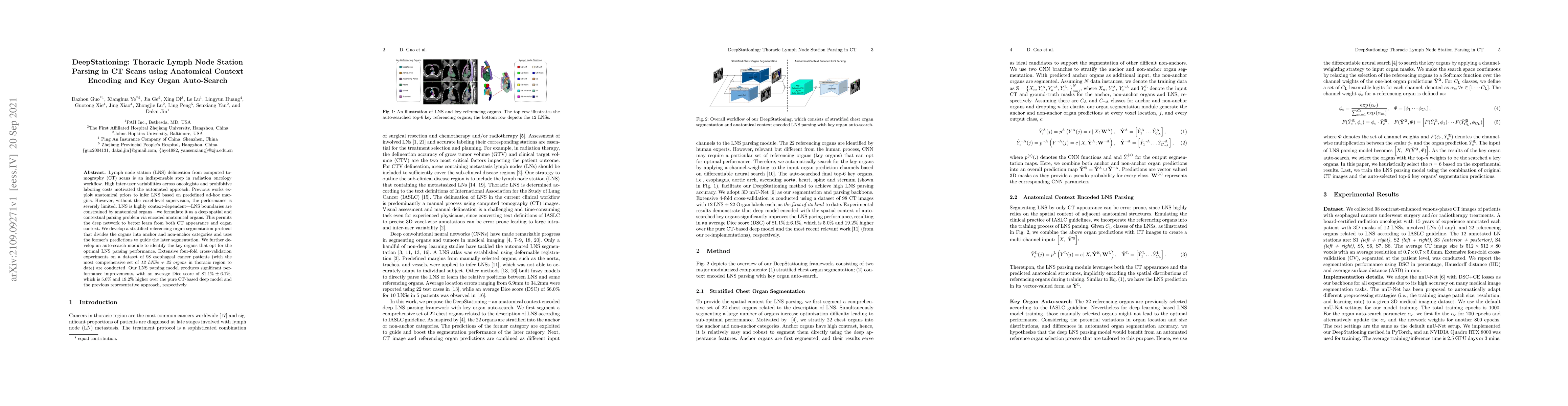
Lymph node station (LNS) delineation from computed tomography (CT) scans is an indispensable step in radiation oncology workflow. High inter-user variabilities across oncologists and prohibitive laboring costs motivated the automated approach. Previous works exploit anatomical priors to infer LNS based on predefined ad-hoc margins. However, without voxel-level supervision, the performance is severely limited. LNS is highly context-dependent - LNS boundaries are constrained by anatomical organs - we formulate it as a deep spatial and contextual parsing problem via encoded anatomical organs. This permits the deep network to better learn from both CT appearance and organ context. We develop a stratified referencing organ segmentation protocol that divides the organs into anchor and non-anchor categories and uses the former's predictions to guide the later segmentation. We further develop an auto-search module to identify the key organs that opt for the optimal LNS parsing performance. Extensive four-fold cross-validation experiments on a dataset of 98 esophageal cancer patients (with the most comprehensive set of 12 LNSs + 22 organs in thoracic region to date) are conducted. Our LNS parsing model produces significant performance improvements, with an average Dice score of 81.1% +/- 6.1%, which is 5.0% and 19.2% higher over the pure CT-based deep model and the previous representative approach, respectively.

Citation & Reference Network Visualization

This graph shows how this paper is connected to other research. The current paper is shown in dark gray, papers that cite it are in green, and papers it references are in blue. You can:

Current Paper
Papers citing this work (Citations)
Papers cited by this work (References)

• Hover over any node to see the paper title

• Click a node to view that paper's details

• Drag nodes to rearrange the graph

• Use the zoom controls (bottom right) to zoom in/out

Citations and References

Title Authors Year Actions
UniReg: Foundation Model for Controllable Medical Image Registration
Zi Li, Jianpeng Zhang, Tai Ma, Tony C. W. Mok, Yan-Jie Zhou, Zeli Chen, X. Ye, Le Lu, Dakai Jin
2025
View Paper
From Slices to Sequences: Autoregressive Tracking Transformer for Cohesive and Consistent 3D Lymph Node Detection in CT Scans
Qinji Yu, Yirui Wang, K. Yan, Dandan Zheng, Dashan Ai, Dazhou Guo, Zhanghexuan Ji, Yanzhou Su, Yun Bian, Na Shen, Xiaowei Ding, Le Lu, X. Ye, Dakai Jin
2025
View Paper
Development and Validation of Thoracic RTOG Organs at Risk Delineation with Deep Learning under Multiple Datasets’ Fusion
X. Ye, Dazhou Guo, Lujun Zhao, Congying Xie, Dandan Zheng, Haihua Yang, Zhixiang Zhu, Xin Sun, Pingping Dong, Huanhuan Li, Weiwei Kong, Jianzhong Cao, Honglei Chen, Juntao Ran, Kai Ren, Hongxin Su, Hao Hu, Cuimeng Tian, Tianlu Wang, Qiang Zeng, Xiao Hu, Ping Peng, Junhua Zhang, Li Zhang, Tingting Zhang, Lue Zhou, Wenchao Guo, Zhanghexuan Ji, Puyang Wang, Hua Zhang, Jiali Liu, Le Lu, Senxiang Yan, Dakai Jin, Feng-Ming Kong
2024
View Paper
Evaluating the Potential of Metastatic Mediastinal Lymph Node Involvement in Non-Small Cell Lung Cancer Patients through Convolutional Neural Networks Analysis of Chest CT
A. P. Reprint, A. Shevtsov, Andrey O. Nefedov, Iaroslav D. Tominin, Piotr K. Yablonskii, Vladislav D. Tominin, P. Gavrilov, Vsevolod M. Malevanniy, Mariya E. Blokhina, Y. Esakov, Z. Tukvadze, V. Gombolevskiy, Yuriy Vasilev, Mariya Dugova, V. Kozlov, V. Chernina, E. Nalivkina, O. Omelyanskaya, M. Belyaev, Roman Reshetnikov, I. Blokhin
2024
View Paper
Towards a Comprehensive, Efficient and Promptable Anatomic Structure Segmentation Model using 3D Whole-body CT Scans
Heng Guo, Jianfeng Zhang, Jiaxing Huang, Tony C. W. Mok, Dazhou Guo, Ke Yan, Le Lu, Dakai Jin, Minfeng Xu
2024
View Paper
SAME++: A Self-supervised Anatomical eMbeddings Enhanced medical image registration framework using stable sampling and regularized transformation
Lin Tian, Zi Li, Fengze Liu, Xiaoyu Bai, Jia Ge, Le Lu, Marc Niethammer, X. Ye, Ke Yan, D. Jin
2023
View Paper
Anatomy-Aware Lymph Node Detection in Chest CT using Implicit Station Stratification
K. Yan, D. Jin, Dazhou Guo, Minfeng Xu, N. Shen, Xianming Hua, X. Ye, Le Lu
2023
View Paper
Continual Segment: Towards a Single, Unified and Non-forgetting Continual Segmentation Model of 143 Whole-body Organs in CT Scans
Zhanghexuan Ji, Dazhou Guo, Puyang Wang, Ke Yan, Le Lu, Minfeng Xu, Qifeng Wang, J. Ge, Mingchen Gao, X. Ye, D. Jin
2023
View Paper
A deep local attention network for pre-operative lymph node metastasis prediction in pancreatic cancer via multiphase CT imaging
Zhilin Zheng, Xu Fang, Jiawen Yao, Mengmeng Zhu, Le Lu, Lingyun Huang, Jing Xiao, Yu Shi, Hong Lu, Jian-Ping Lu, Ling Zhang, C. Shao, Yun Bian
2023
View Paper
Comprehensive and clinically accurate head and neck cancer organs-at-risk delineation on a multi-institutional study
X. Ye, Dazhou Guo, J. Ge, S. Yan, Yinping Xin, Yuchen Song, Yongheng Yan, Bing-Shen Huang, T. Hung, Zhuotun Zhu, Ling Peng, Yanping Ren, Rui Liu, Gong Zhang, Mengyuan Mao, Xiaohua Chen, Zhongjie Lu, Wenxiang Li, Yuzhen Chen, Lingyun Huang, Jing Xiao, Adam P. Harrison, Le Lu, C. Lin, D. Jin, T. Ho
2022
View Paper
Towards automated organs at risk and target volumes contouring: Defining precision radiation therapy in the modern era
D. Jin, Dazhou Guo, J. Ge, X. Ye, Le Lu
2022
View Paper
Integrating features from lymph node stations for metastatic lymph node detection
Chaoyi Wu, Feng Chang, Xiao Su, Zhihan Wu, Yanfeng Wang, Ling Zhu, Ya Zhang
2022
View Paper
Multi-Institutional Validation of Two-Streamed Deep Learning Method for Automated Delineation of Esophageal Gross Tumor Volume Using Planning CT and FDG-PET/CT
X. Ye, Dazhou Guo, C. Tseng, J. Ge, T. Hung, P. Pai, Yanping Ren, Lu Zheng, Xinli Zhu, Ling Peng, Ying Chen, Xiaohua Chen, C-Y Chou, Danni Chen, Jiaze Yu, Yuzhen Chen, Feiran Jiao, Yinping Xin, Lingyun Huang, G. Xie, Jing Xiao, Le Lu, Senxiang Yan, D. Jin, T. Ho
2021
View Paper
SAME: Deformable Image Registration Based on Self-supervised Anatomical Embeddings
Fengze Liu, K. Yan, Adam P. Harrison, Dazhou Guo, Le Lu, A. Yuille, Lingyun Huang, G. Xie, Jing Xiao, X. Ye, D. Jin
2021
View Paper
Automated Coarse-to-fine Segmentation of Thoracic Duct using Anatomy Priors and Topology-guided Curved Planar Reformation
Puyang Wang, Panwen Hu, Jiali Liu, Hang Yu, X. Ye, Jinliang Zhang, Hui Li, Li Yang, Le Lu, D. Jin, Feng-Ming Kong
2023
View Paper
Thoracic Lymph Node Segmentation in CT Imaging via Lymph Node Station Stratification and Size Encoding
Dazhou Guo, J. Ge, K. Yan, Puyang Wang, Zhuotun Zhu, D. Zheng, Xiansheng Hua, Le Lu, T. Ho, X. Ye, D. Jin
2022
View Paper
Medical Image Analysis
Lin Tian, Zi Li, Fengze Liu, Xiaoyu Bai, Jia Ge, Le Lu, Marc Niethammer, X. Ye, Ke Yan, D. Jin
View Paper
Automatic Delineation of Thoracic RTOG Organs at Risk with A Deep Learning Model
X. Ye, Dazhou Guo, Puyang Wang, Zhanghexuan Ji, Dandan Zheng, Jiali Liu, Honglei Chen, Pingping Dong, Weiwei Kong, Huanhuan Li, Juntao Ran, Kai Ren, Hongxin Su, Xin Sun, Congying Xie, Hao Hu, Cuimeng Tian, Tianlu Wang, Haihua Yang, Qiang Zeng, Zhixiang Zhu, Jianzhong Cao, Xiao Hu, Ping Peng, Junhua Zhang, Li Zhang, Tingting Zhang, Lue Zhou, Wenchao Guo, Hua Zhang, Le Lu, Lujun Zhao, Dakai Jin, Spring Kong, Hua Zhang aa
View Paper
Computerized Medical Imaging and Graphics
Rui Zhang, Lin Huang, W. Xia, Bo Zhang, B. Qiu, Xin Gao
View Paper

Similar Papers

Anatomy-Aware Lymph Node Detection in Chest CT using Implicit Station Stratification

Xian-Sheng Hua, Le Lu, Ke Yan, Minfeng Xu, Xianghua Ye, Dazhou Guo, Na Shen, Dakai Jin

Published: 2023-07-31

Segmentation of Mediastinal Lymph Nodes in CT with Anatomical Priors

Ronald M. Summers, Tejas Sudharshan Mathai, Bohan Liu

Published: 2024-01-15

SDF-Net: A Hybrid Detection Network for Mediastinal Lymph Node Detection on Contrast CT Images

Dinggang Shen, Lanzhuju Mei, Jiameng Liu, Jiuli Xiong, Zhong Xue, Xiaohuan Cao

Published: 2024-09-10

LNQ Challenge 2023: Learning Mediastinal Lymph Node Segmentation with a Probabilistic Lymph Node Atlas

Joshua Niemeijer, Jan Ehrhardt, Heinz Handels, Sofija Engelson, Timo Kepp

Published: 2024-06-07

Comments (0)

This website uses cookies to ensure you get the best experience. Learn more