Publication
Metrics
AI Quick Summary
This paper uses a continuum model to explore the physical mechanisms behind cell division by comparing it to nematic liquid crystal droplets. It highlights the role of microtubule assembly and cell cortex elasticity in shaping cell division, predicting a critical parameter value for successful division and suggesting that microtubule anchoring at the cortex is crucial for bipolar division.
Paper Preview
Abstract
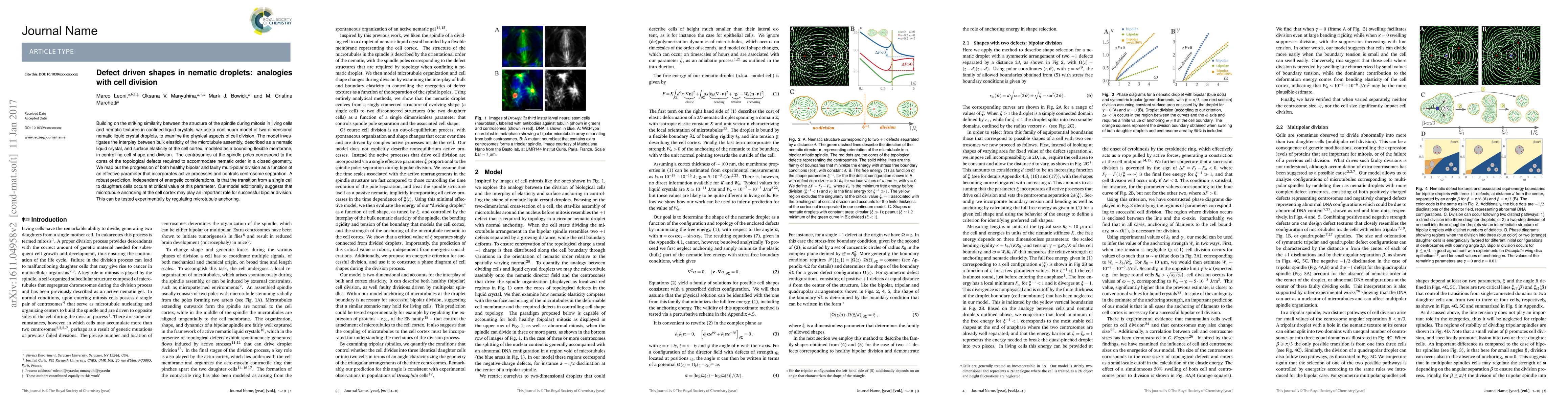
Building on the striking similarity between the structure of the spindle during mitosis in living cells and nematic textures in confined liquid crystals, we use a continuum model of two-dimensional nematic liquid crystal droplets, to examine the physical aspects of cell division. The model investigates the interplay between bulk elasticity of the microtubule assembly, described as a nematic liquid crystal, and surface elasticity of the cell cortex, modelled as a bounding flexible membrane, in controlling cell shape and division. The centrosomes at the spindle poles correspond to the cores of the topological defects required to accommodate nematic order in a closed geometry. We map out the progression of both healthy bipolar and faulty multi-polar division as a function of an effective parameter that incorporates active processes and controls centrosome separation. A robust prediction, independent of energetic considerations, is that the transition from a single cell to daughters cells occurs at critical value of this parameter. Our model additionally suggests that microtubule anchoring at the cell cortex may play an important role for successful bipolar division. This can be tested experimentally by regulating microtubule anchoring.
AI Key Findings
Get AI-generated insights about this paper's methodology, results, significance, and more — seven facets brought into focus.
Impact
Paper Details
PDF Preview
Key Terms
Citation Network
Current paper (gray), citations (green), references (blue)
Display is limited for performance on very large graphs.
Discussion 0