Summary
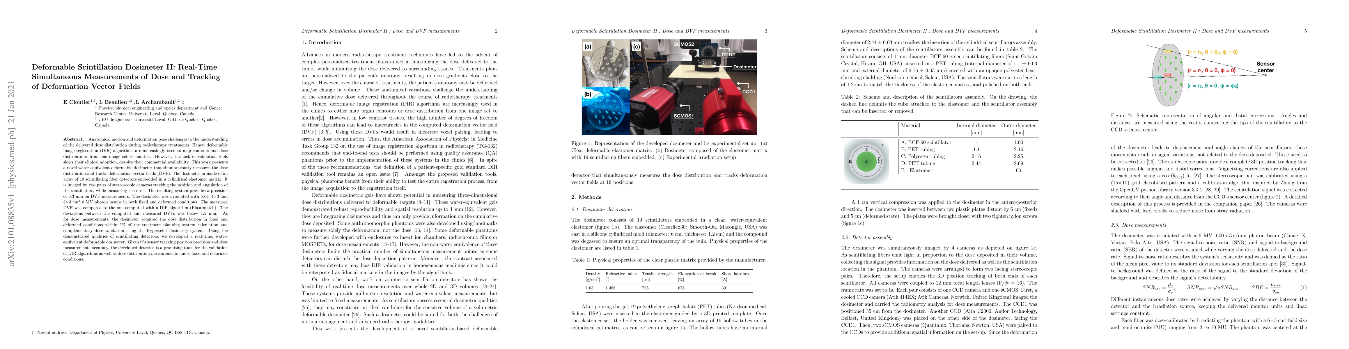
Anatomical motion and deformation pose challenges to the understanding of the delivered dose distribution during radiotherapy treatments. Hence, deformable image registration (DIR) algorithms are increasingly used to map contours and dose distributions from one image set to another. However, the lack of validation tools slows their clinical adoption, despite their commercial availability. This work presents a novel water-equivalent deformable dosimeter that simultaneously measures the dose distribution and tracks deformation vector fields (DVF). The dosimeter in made of an array of 19 scintillating fiber detectors embedded in a cylindrical elastomer matrix. It is imaged by two pairs of stereoscopic cameras tracking the position and angulation of the scintillators, while measuring the dose. The resulting system provides a precision of 0.3 mm on DVF measurements. The dosimeter was irradiated with 5$\times$3, 4$\times$3 and 3$\times$3 cm$^2$ 6 MV photon beams in both fixed and deformed conditions. The measured DVF was compared to the one computed with a DIR algorithm (Plastimatch). The deviations between the computed and measured DVFs was below 1.5 mm. As for dose measurements, the dosimeter acquired the dose distribution in fixed and deformed conditions within 1\% of the treatment planning system calculation and complementary dose validation using the Hyperscint dosimetry system. Using the demonstrated qualities of scintillating detectors, we developed a real-time, water-equivalent deformable dosimeter. Given it's sensor tracking position precision and dose measurements accuracy, the developed detector is a promising tools for the validation of DIR algorithms as well as dose distribution measurements under fixed and deformed conditions.
AI Key Findings
Get AI-generated insights about this paper's methodology, results, and significance.
Paper Details
PDF Preview
Key Terms
Citation Network
Current paper (gray), citations (green), references (blue)
Display is limited for performance on very large graphs.
Similar Papers
Found 4 papersDeformable Scintillation Dosimeter I: Challenges and Implementation using Computer Vision Techniques
Quantitative real-time measurements of dose and dose rate in UHDR proton pencil beams via scintillation imaging system
Megan Clark, Joseph Harms, Roman Vasyltsiv et al.
| Title | Authors | Year | Actions |
|---|
Comments (0)