ArxivLens

Quick Summary:

This paper proposes an improved method for segmenting and classifying dermatoscopic skin lesion images using U-Net for segmentation and DenseNet121 for classification. The combined approach shows promising results on the ISIC-2018 and HAM10000 datasets, achieving high accuracy in distinguishing cancerous and non-cancerous samples.

arXiv Id: 2110.04632
Date Published: 2022-02-17
Date Updated: 2022-02-17

Categories:

Key Terms (TF-IDF):

skin (0.297)
cancer (0.289)
cancer (0.269)
segmentation (0.250)
classification (0.240)
classification (0.233)
image (0.230)
segmentation (0.228)
image (0.217)
net (0.203)
architectures (0.198)
steps (0.195)
net (0.191)
architectures (0.181)
steps (0.178)
accuracy (0.173)
accuracy (0.165)
samples (0.160)
samples (0.156)
dataset (0.153)
License: http://creativecommons.org/licenses/by/4.0/

Summary:

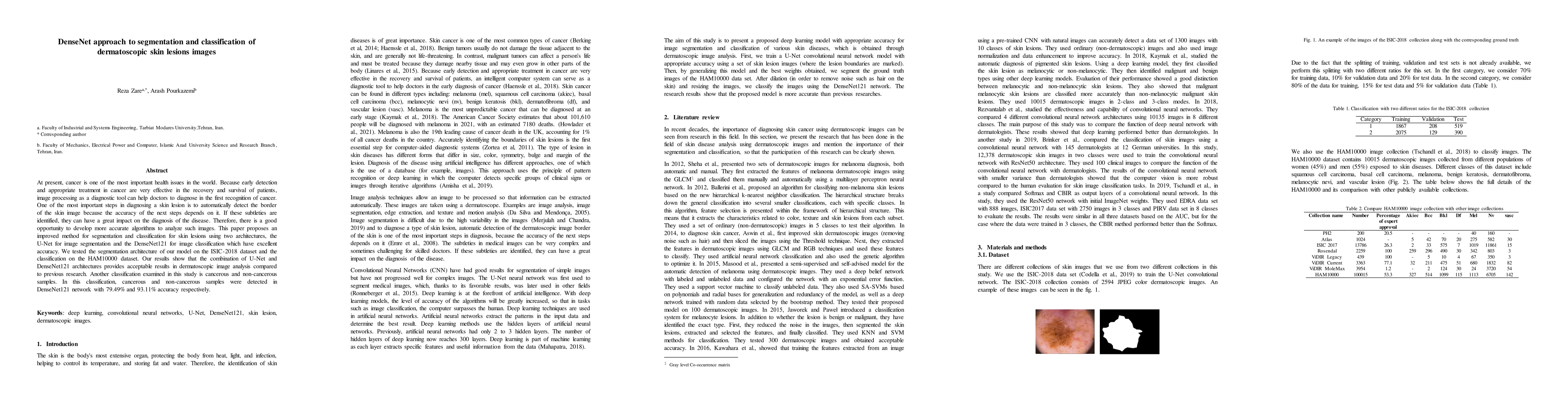
At present, cancer is one of the most important health issues in the world. Because early detection and appropriate treatment in cancer are very effective in the recovery and survival of patients, image processing as a diagnostic tool can help doctors to diagnose in the first recognition of cancer. One of the most important steps in diagnosing a skin lesion is to automatically detect the border of the skin image because the accuracy of the next steps depends on it. If these subtleties are identified, they can have a great impact on the diagnosis of the disease. Therefore, there is a good opportunity to develop more accurate algorithms to analyze such images. This paper proposes an improved method for segmentation and classification for skin lesions using two architectures, the U-Net for image segmentation and the DenseNet121 for image classification which have excellent accuracy. We tested the segmentation architecture of our model on the ISIC-2018 dataset and the classification on the HAM10000 dataset. Our results show that the combination of U-Net and DenseNet121 architectures provides acceptable results in dermatoscopic image analysis compared to previous research. Another classification examined in this study is cancerous and non-cancerous samples. In this classification, cancerous and non-cancerous samples were detected in DenseNet121 network with 79.49% and 93.11% accuracy respectively.

Citation & Reference Network Visualization

This graph shows how this paper is connected to other research. The current paper is shown in dark gray, papers that cite it are in green, and papers it references are in blue. You can:

Current Paper
Papers citing this work (Citations)
Papers cited by this work (References)

• Hover over any node to see the paper title

• Click a node to view that paper's details

• Drag nodes to rearrange the graph

• Use the zoom controls (bottom right) to zoom in/out

Citations and References

Title Authors Year Actions
Application of Deep Learning Convolutional Neural Network for Melanoma Skin Cancer Classification
Hamdi Syukron, Mustakim
2024
View Paper
SkinSage - Lesion Diagnosis Using Deep Learning Techniques
Geetika, Paarth Bhardwaj, Nimish, Vaibhav Bhargava, Shivendra Singh
2024
View Paper
Diagnosing Melanomas in Dermoscopy Images Using Deep Learning
G. Alwakid, Walaa Gouda, M. Humayun, Noor Zaman Jhanjhi
2023
View Paper
Melanoma Malignancy Prognosis Using Deep Transfer Learning
R. Shobarani, R. Sharmila, M. Kathiravan, A. A. Pandian, Ch Narasimha Chary, K. Vigneshwaran
2023
View Paper
Melanoma Detection Using Deep Learning-Based Classifications
G. Alwakid, Walaa Gouda, M. Humayun, N. Sama
2022
View Paper

Comments (0)

This website uses cookies to ensure you get the best experience. Learn more