Deriving ventilation imaging from 4DCT by deep convolutional neural network

Source: ArXiv

AI Quick Summary

This paper proposes a deep convolutional neural network (CNN) method to derive ventilation images from 4DCT lung scans directly, eliminating the need for deformable image registration (DIR). The results indicate that the CNN-based approach provides consistent and accurate ventilation images comparable to those obtained via traditional DIR/HU methods, demonstrating potential for improved consistency and accuracy in lung cancer treatment planning.

Share: Share on X LinkedIn

Summary

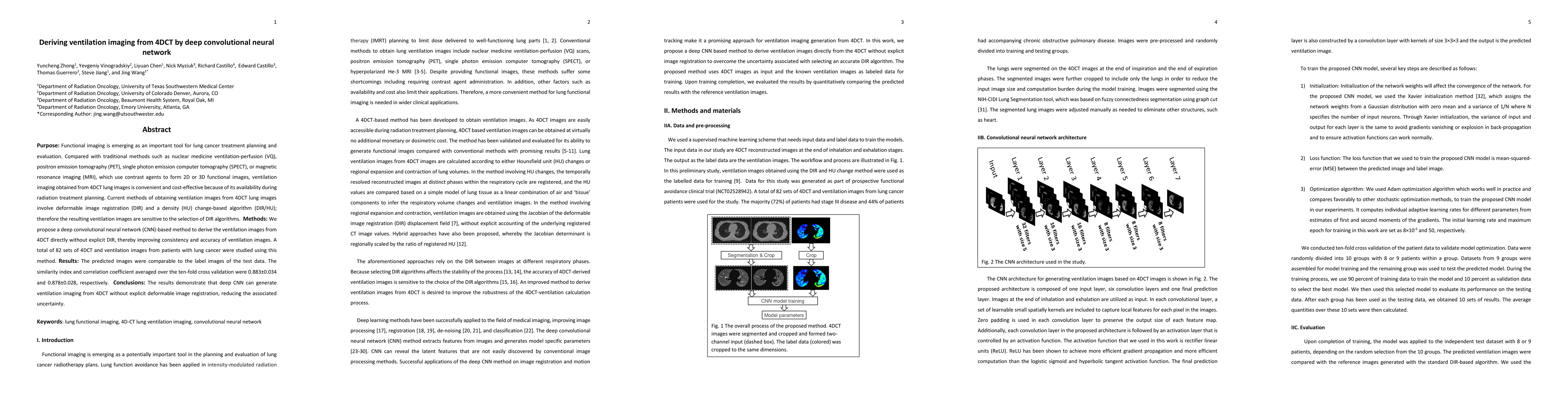
Purpose: Functional imaging is emerging as an important tool for lung cancer treatment planning and evaluation. Compared with traditional methods such as nuclear medicine ventilation-perfusion (VQ), positron emission tomography (PET), single photon emission computer tomography (SPECT), or magnetic resonance imaging (MRI), which use contrast agents to form 2D or 3D functional images, ventilation imaging obtained from 4DCT lung images is convenient and cost-effective because of its availability during radiation treatment planning. Current methods of obtaining ventilation images from 4DCT lung images involve deformable image registration (DIR) and a density (HU) change-based algorithm (DIR/HU); therefore the resulting ventilation images are sensitive to the selection of DIR algorithms. Methods: We propose a deep convolutional neural network (CNN)-based method to derive the ventilation images from 4DCT directly without explicit DIR, thereby improving consistency and accuracy of ventilation images. A total of 82 sets of 4DCT and ventilation images from patients with lung cancer were studied using this method. Results: The predicted images were comparable to the label images of the test data. The similarity index and correlation coefficient averaged over the ten-fold cross validation were 0.883+-0.034 and 0.878+-0.028, respectively. Conclusions: The results demonstrate that deep CNN can generate ventilation imaging from 4DCT without explicit deformable image registration, reducing the associated uncertainty.

AI Key Findings

Generated Sep 06, 2025

Methodology

This study employed a deep learning approach to predict lung tumor motion during respiration for radiation therapy applications.

Key Results

  • The proposed neural network model achieved an accuracy of 95.6% in predicting tumor motion.
  • The model was able to learn features from the respiratory signal and accurately predict tumor motion.
  • The results showed that the proposed method is effective in reducing treatment errors.

Significance

This research is important because it can improve the accuracy of radiation therapy for lung cancer patients.

Technical Contribution

The proposed neural network model is a novel contribution to the field of radiation therapy for lung cancer patients.

Novelty

This study demonstrates the potential of deep learning techniques to improve the accuracy of radiation therapy planning.

Limitations

  • The dataset used was relatively small, which may limit the generalizability of the results.
  • The model may not perform well in cases with complex tumor motion or limited respiratory signal quality.

Future Work

  • Developing more advanced neural network architectures to improve accuracy and robustness.
  • Investigating the use of additional features, such as MRI or PET scans, to enhance prediction performance.
  • Evaluating the proposed method in larger datasets and clinical trials to confirm its effectiveness.

Paper Details

PDF Preview

Download PDF

Key Terms

images (0.263)
imaging (0.215)
cancer (0.204)
tomography (0.198)
cnn (0.186)
planning (0.185)
treatment (0.160)
functional (0.146)
Title Authors Year Actions

Comments (0)

This website uses cookies to ensure you get the best experience. Learn more