Quick Summary:

This paper describes the design of a 3000-pixel transition-edge sensor x-ray spectrometer for microcircuit tomography, combining a scanning electron microscope with a TES x-ray spectrometer to non-destructively image complex integrated circuits. The upgrade aims to enhance imaging speed by up to 60 times through increased detector count and speed.

Summary

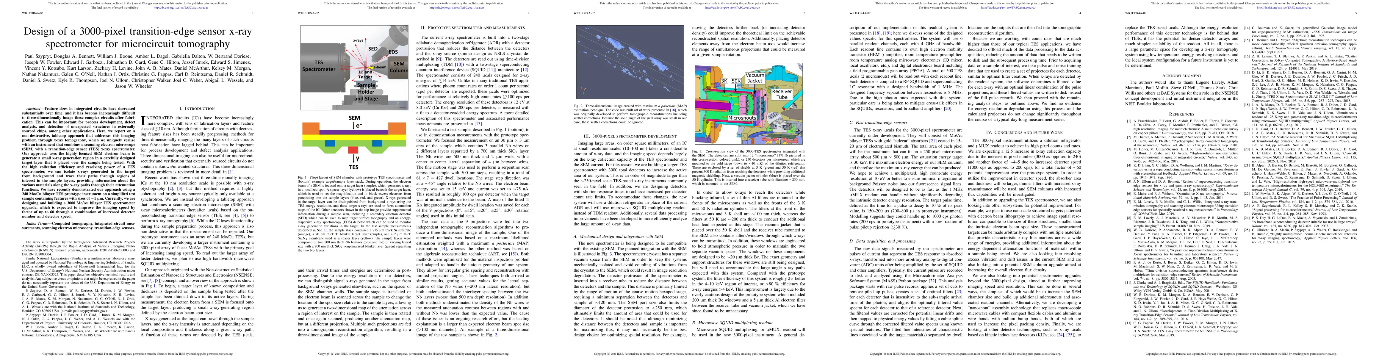
Feature sizes in integrated circuits have decreased substantially over time, and it has become increasingly difficult to three-dimensionally image these complex circuits after fabrication. This can be important for process development, defect analysis, and detection of unexpected structures in externally sourced chips, among other applications. Here, we report on a non-destructive, tabletop approach that addresses this imaging problem through x-ray tomography, which we uniquely realize with an instrument that combines a scanning electron microscope (SEM) with a transition-edge sensor (TES) x-ray spectrometer. Our approach uses the highly focused SEM electron beam to generate a small x-ray generation region in a carefully designed target layer that is placed over the sample being tested. With the high collection efficiency and resolving power of a TES spectrometer, we can isolate x-rays generated in the target from background and trace their paths through regions of interest in the sample layers, providing information about the various materials along the x-ray paths through their attenuation functions. We have recently demonstrated our approach using a 240 Mo/Cu bilayer TES prototype instrument on a simplified test sample containing features with sizes of $\sim$1 $\mu$m. Currently, we are designing and building a 3000 Mo/Au bilayer TES spectrometer upgrade, which is expected to improve the imaging speed by factor of up to 60 through a combination of increased detector number and detector speed.

AI Key Findings

Get AI-generated insights about this paper's methodology, results, and significance.

Paper Details

PDF Preview

Download PDF

Key Terms

spectrometer (0.247)
spectrometer (0.240)
mo (0.210)
bilayer (0.208)
circuits (0.197)
bilayer (0.194)
instrument (0.184)
instrument (0.182)
ray (0.180)
circuits (0.178)
ray (0.172)
paths (0.169)
paths (0.168)
sizes (0.160)
detector (0.155)
sizes (0.152)
sample (0.151)
detector (0.150)
sample (0.149)
speed (0.146)

Citation Network

Current paper (gray), citations (green), references (blue)

Current Paper
Citations
References
Click to view
Title Authors Year Actions
A High-Speed, High-Resolution Transition Edge Sensor Spectrometer for Soft X-Rays at the Advanced Photon Source
O. Quaranta, Don Jensen, Kelsey Morgan, Joel Weber, J. McChesney, Hao Zheng, T. Guruswamy, Jonathan Baldwin, Ben Mates, Nathan Ortiz, J. Gard, Doug Bennet, Dan Schmidt, L. Gades, Antonino Miceli
2025
View Paper
Low-noise cryogenic microwave amplifier characterization with a calibrated noise source.
M. Malnou, T. F. Q. Larson, J. D. Teufel, F. Lecocq, J. Aumentado
2023
View Paper
Nanoscale X-Ray Tomography of Integrated Circuits Using a Hybrid Electron/X-Ray Microscope: Results and Prospects
Nathan Nakamura, P. Szypryt, J. W. Fowler, Zachary H. Levine, D. Swetz
2023
View Paper
A tabletop X-ray tomography instrument for nanometer-scale imaging: reconstructions
Z. Levine, B. Alpert, Amber L. Dagel, J. Fowler, Edward S. Jimenez, Nathan Nakamura, D. Swetz, P. Szypryt, K. Thompson, J. Ullom
2023
View Paper
Improved microwave SQUID multiplexer readout using a kinetic-inductance traveling-wave parametric amplifier
M. Malnou, J. Mates, M. Vissers, L. Vale, D. R. Schmidt, D. Bennett, J. Gao, J. Ullom
2023
View Paper
A Tabletop X-Ray Tomography Instrument for Nanometer-Scale Imaging: Demonstration of the 1,000-Element Transition-Edge Sensor Subarray
P. Szypryt, Nathan Nakamura, D. Becker, D. Bennett, Amber L. Dagel, W. Doriese, J. Fowler, J. Gard, J. Harris, G. Hilton, J. Imrek, Edward S. Jimenez, K. Larson, Z. Levine, J. Mates, D. McArthur, L. Miaja-Avila, K. Morgan, G. O’Neil, N. Ortiz, C. Pappas, D. Schmidt, K. Thompson, J. Ullom, L. Vale, M. Vissers, C. Walker, J. Weber, A. Wessels, Jason W. Wheeler, D. Swetz
2022
View Paper
Nanoscale Three-Dimensional Imaging of Integrated Circuits Using a Scanning Electron Microscope and Transition-Edge Sensor Spectrometer
Nathan Nakamura, P. Szypryt, Amber L. Dagel, B. Alpert, D. Bennett, W. Doriese, M. Durkin, J. Fowler, D. Fox, J. Gard, Ryan N. Goodner, James Zachariah Harris, G. Hilton, Edward S. Jimenez, Burke L. Kernen, K. Larson, Z. Levine, D. McArthur, K. Morgan, G. O’Neil, N. Ortiz, C. Pappas, C. Reintsema, D. Schmidt, Peter A. Schultz, K. Thompson, J. Ullom, L. Vale, C. Vaughan, C. Walker, J. Weber, Jason W. Wheeler, D. Swetz
2022
View Paper
Signal readout for Transition-Edge Sensor X-ray imaging spectrometers
H. Akamatsu, W. Doriese, J. Mates, B. Jackson
2022
View Paper
Physics-assisted Generative Adversarial Network for X-Ray Tomography
Zhen Guo, J. Song, G. Barbastathis, M. Glinsky, C. Vaughan, K. Larson, B. Alpert, Z. Levine
2022
View Paper
Transition edge sensor-based detector: from X-ray to $$\gamma$$-ray
Shuo Zhang, Jingkai Xia, Tao Sun, Wendou Wu, Bin Wu, Yong-Liang Wang, R. Cantor, K. Han, Xiao-Peng Zhou, Hao-Ran Liu, Fuqing Fan, Siming Guo, Juncheng Liang, Dengwan Li, Yanping Song, X. Ju, Qiangzhi Fu, Zhi Liu
2022
View Paper
Advantage of Machine Learning over Maximum Likelihood in Limited-Angle Low-Photon X-Ray Tomography
Zhen Guo, J. Song, G. Barbastathis, M. Glinsky, C. Vaughan, K. Larson, B. Alpert, Z. Levine
2021
View Paper
Transition edge sensor-based detector: from X-ray to c -ray
Shuo Zhang, Jingkai Xia, Tao Sun, Wenjuan Wu, Bin Wu, Yong-Liang Wang, R. Cantor, K. Han, Xiao-Peng Zhou, Hao-Ran Liu, Fuqing Fan, Siming Guo, Juncheng Liang, Dengwan Li, Yanping Song, X. Ju, Qiangzhi Fu, Zhi Liu
2022
View Paper

Comments (0)

This website uses cookies to ensure you get the best experience. Learn more