Detecting Early Kidney Allograft Fibrosis with Multi-b-value Spectral Diffusion MRI
Publication
Metrics
Paper Preview
Abstract
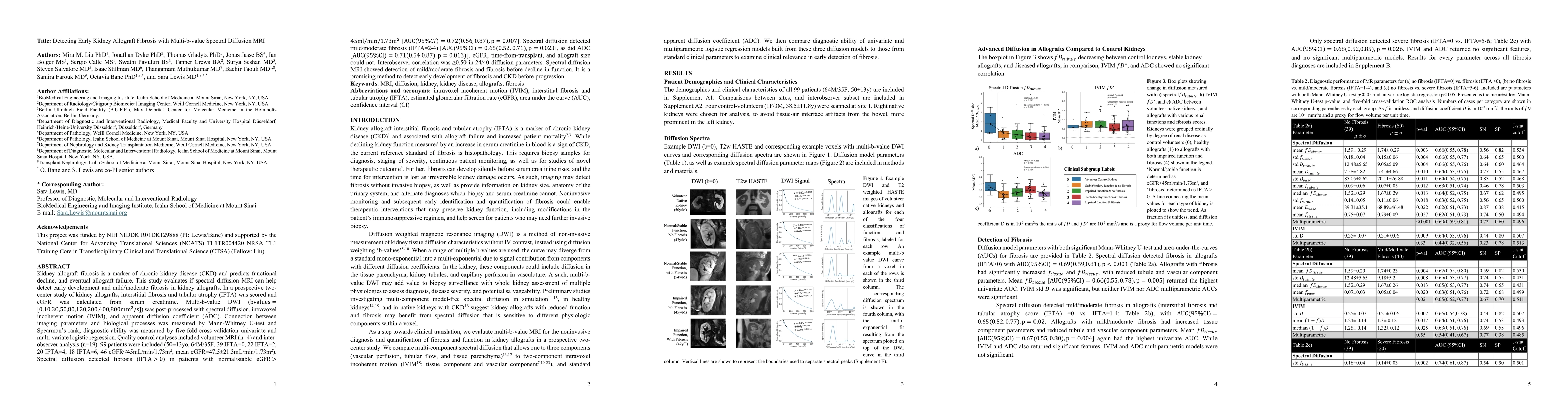
Kidney allograft fibrosis is a marker of chronic kidney disease (CKD) and predicts functional decline, and eventual allograft failure. This study evaluates if spectral diffusion MRI can help detect early development and mild/moderate fibrosis in kidney allografts. In a prospective two-center study of kidney allografts, interstitial fibrosis and tubular atrophy (IFTA) was scored and eGFR was calculated from serum creatinine. Multi-b-value DWI (bvalues=[0,10,30,50,80,120,200,400,800mm2/s]) was post-processed with spectral diffusion, intravoxel incoherent motion (IVIM), and apparent diffusion coefficient (ADC). Connection between imaging parameters and biological processes was measured by Mann-Whitney U-test and Spearman's rank; diagnostic ability was measured by five-fold cross-validation univariate and multi-variate logistic regression. Quality control analyses included volunteer MRI (n=4) and inter-observer analysis (n=19). 99 patients were included (50$\pm$13yo, 64M/35F, 39 IFTA=0, 22 IFTA=2, 20 IFTA=4, 18 IFTA=6, 46 eGFR<=45mL/min/1.73m2, mean eGFR=47.5$\pm$21.3mL/min/1.73m2). Spectral diffusion detected fibrosis (IFTA>0) in patients with normal/stable eGFR>45ml/min/1.73m2 [AUC(95$\%$CI)=0.72(0.56,0.87),p=0.007]. Spectral diffusion detected mild/moderate fibrosis (IFTA=2-4) [AUC(95$\%$CI)=0.65(0.52,0.71),p=0.023], as did ADC [AUC(95$\%$CI)=0.71(0.54,0.87),p=0.013)]. eGFR, time-from-transplant, and allograft size could not. Interobserver correlation was >0.50 in 24 out of 40 diffusion parameters. Spectral diffusion MRI showed detection of mild/moderate fibrosis and fibrosis before decline in function. It is a promising method to detect early development of fibrosis and CKD before progression.
AI Key Findings
Get AI-generated insights about this paper's methodology, results, significance, and more — seven facets brought into focus.
Discussion 0