Detection-aided liver lesion segmentation using deep learning
Publication
Metrics
Paper Preview
Abstract
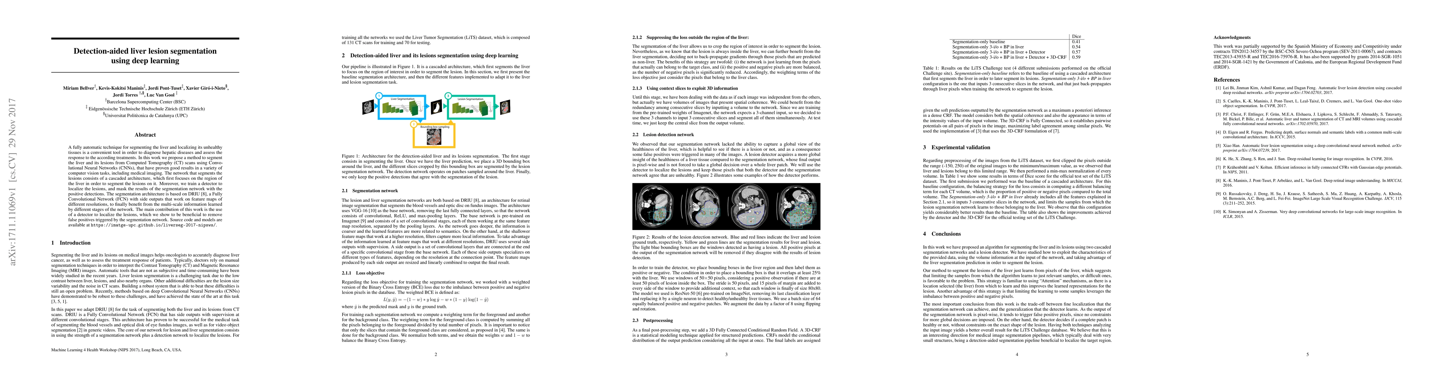
A fully automatic technique for segmenting the liver and localizing its unhealthy tissues is a convenient tool in order to diagnose hepatic diseases and assess the response to the according treatments. In this work we propose a method to segment the liver and its lesions from Computed Tomography (CT) scans using Convolutional Neural Networks (CNNs), that have proven good results in a variety of computer vision tasks, including medical imaging. The network that segments the lesions consists of a cascaded architecture, which first focuses on the region of the liver in order to segment the lesions on it. Moreover, we train a detector to localize the lesions, and mask the results of the segmentation network with the positive detections. The segmentation architecture is based on DRIU, a Fully Convolutional Network (FCN) with side outputs that work on feature maps of different resolutions, to finally benefit from the multi-scale information learned by different stages of the network. The main contribution of this work is the use of a detector to localize the lesions, which we show to be beneficial to remove false positives triggered by the segmentation network. Source code and models are available at https://imatge-upc.github.io/liverseg-2017-nipsws/ .
AI Key Findings
Get AI-generated insights about this paper's methodology, results, significance, and more — seven facets brought into focus.
Impact
Paper Details
PDF Preview
Key Terms
Citation Network
Current paper (gray), citations (green), references (blue)
Display is limited for performance on very large graphs.
Discussion 0