Summary
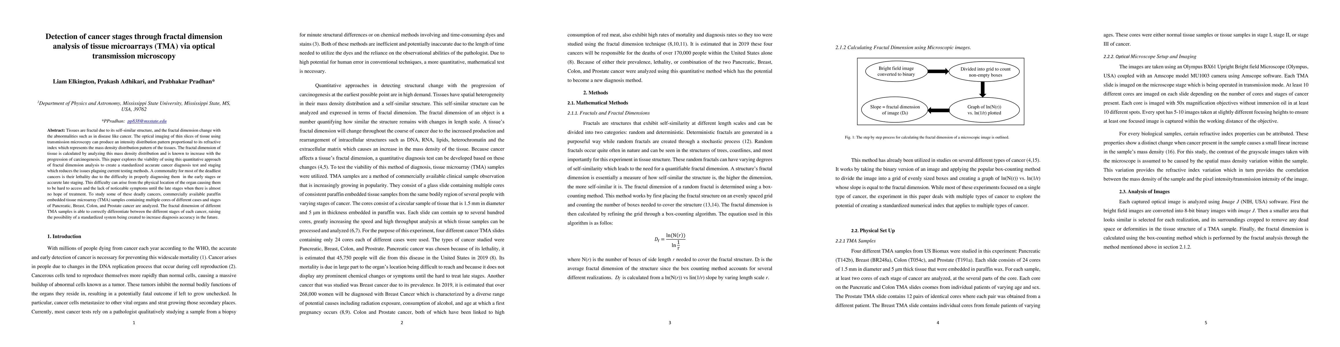
Tissues are fractal due to its self-similar structure, and the fractal dimension change with the abnormalities such as in disease like cancer. The optical imaging of thin slices of tissue using transmission microscopy can produce an intensity distribution pattern proportional to its refractive index which represents the mass density distribution pattern of the tissues. The fractal dimension of tissue is calculated by analyzing this mass density distribution and is known to increase with the progression of carcinogenesis. This paper explores the viability of using this quantitative approach of fractal dimension analysis to create a standardized accurate cancer diagnosis test and staging which reduces the issues plaguing current testing methods. A commonality for most of the deadliest cancers is their lethality due to the difficulty in properly diagnosing them in the early stages or accurate late staging. This difficulty can arise from the physical location of the organ causing them to be hard to access and the lack of noticeable symptoms until the late stages when there is almost no hope of treatment. To study some of these deadly cancers, commercially available paraffin embedded tissue microarray (TMA) samples containing multiple cores of different cases and stages of Pancreatic, Breast, Colon, and Prostate cancer are analyzed. The fractal dimension of different TMA samples is able to correctly differentiate between the different stages of each cancer, raising the possibility of a standardized system being created to increase diagnosis accuracy in the future.
AI Key Findings
Get AI-generated insights about this paper's methodology, results, and significance.
Paper Details
PDF Preview
Key Terms
Citation Network
Current paper (gray), citations (green), references (blue)
Display is limited for performance on very large graphs.
Similar Papers
Found 4 papersNo citations found for this paper.
Comments (0)