Summary
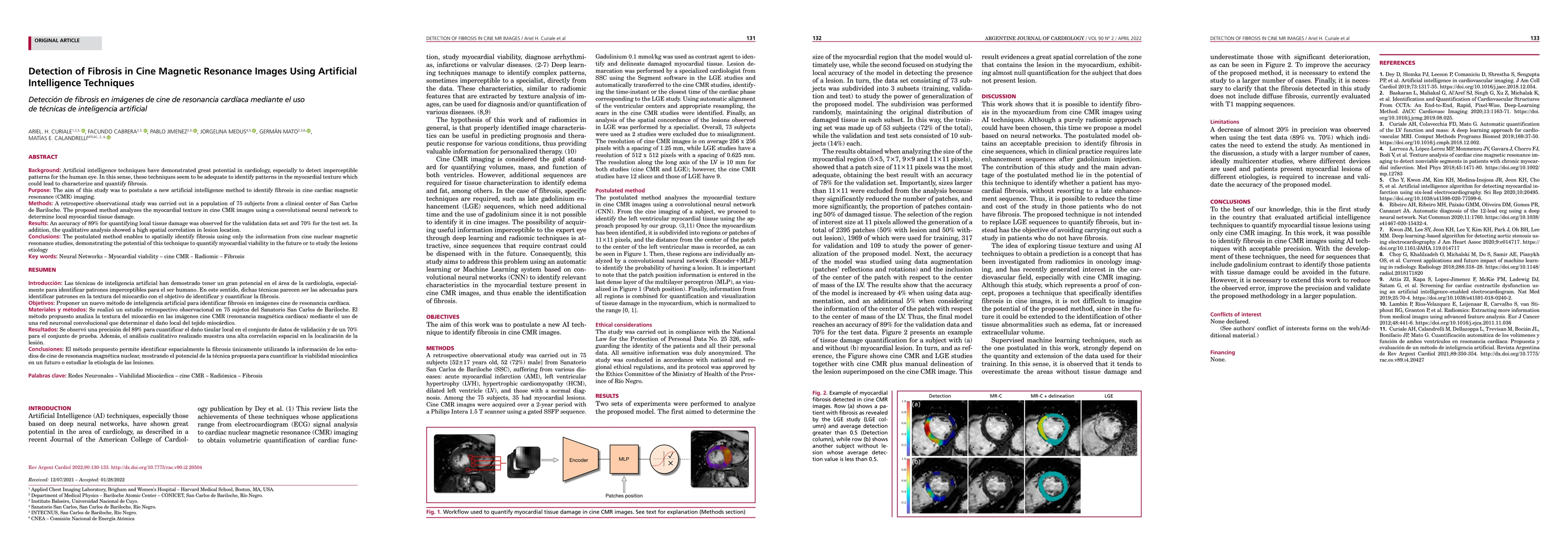
Background: Artificial intelligence techniques have demonstrated great potential in cardiology, especially to detect imperceptible patterns for the human eye. In this sense, these techniques seem to be adequate to identify patterns in the myocardial texture which could lead to characterize and quantify fibrosis. Purpose: The aim of this study was to postulate a new artificial intelligence method to identify fibrosis in cine cardiac magnetic resonance (CMR) imaging. Methods: A retrospective observational study was carried out in a population of 75 subjects from a clinical center of San Carlos de Bariloche. The proposed method analyzes the myocardial texture in cine CMR images using a convolutional neural network to determine local myocardial tissue damage. Results: An accuracy of 89% for quantifying local tissue damage was observed for the validation data set and 70% for the test set. In addition, the qualitative analysis showed a high spatial correlation in lesion location. Conclusions: The postulated method enables to spatially identify fibrosis using only the information from cine nuclear magnetic resonance studies, demonstrating the potential of this technique to quantify myocardial viability in the future or to study the lesions etiology
AI Key Findings
Get AI-generated insights about this paper's methodology, results, and significance.
Paper Details
PDF Preview
Key Terms
Citation Network
Current paper (gray), citations (green), references (blue)
Display is limited for performance on very large graphs.
Similar Papers
Found 4 papersBeyond traditional Magnetic Resonance processing with Artificial Intelligence
Amir Jahangiri, Vladislav Orekhov
Fully Automated Assessment of Cardiac Coverage in Cine Cardiovascular Magnetic Resonance Images using an Explainable Deep Visual Salient Region Detection Model
Mohammad Hashemi, Shahabedin Nabavi, Mohsen Ebrahimi Moghaddam et al.
Malware Detection and Prevention using Artificial Intelligence Techniques
Fan Wu, Akond Rahman, Hossain Shahriar et al.
No citations found for this paper.

Comments (0)