Summary
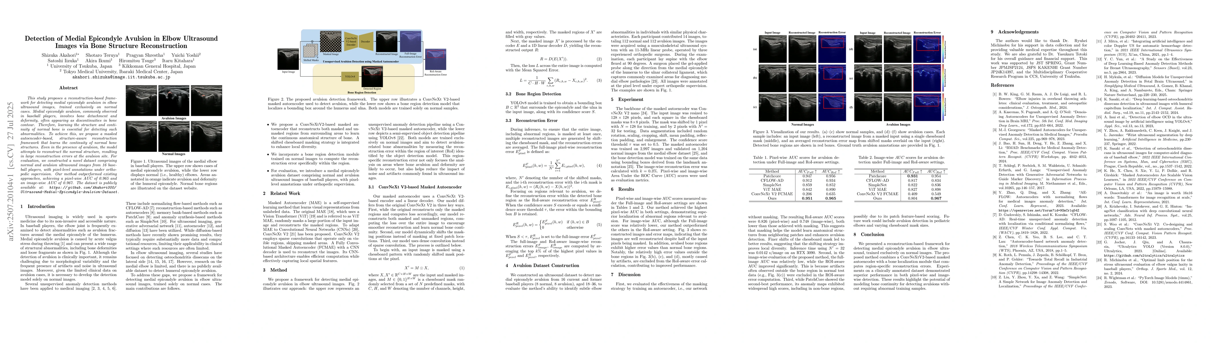
This study proposes a reconstruction-based framework for detecting medial epicondyle avulsion in elbow ultrasound images, trained exclusively on normal cases. Medial epicondyle avulsion, commonly observed in baseball players, involves bone detachment and deformity, often appearing as discontinuities in bone contour. Therefore, learning the structure and continuity of normal bone is essential for detecting such abnormalities. To achieve this, we propose a masked autoencoder-based, structure-aware reconstruction framework that learns the continuity of normal bone structures. Even in the presence of avulsion, the model attempts to reconstruct the normal structure, resulting in large reconstruction errors at the avulsion site. For evaluation, we constructed a novel dataset comprising normal and avulsion ultrasound images from 16 baseball players, with pixel-level annotations under orthopedic supervision. Our method outperformed existing approaches, achieving a pixel-wise AUC of 0.965 and an image-wise AUC of 0.967. The dataset is publicly available at: https://github.com/Akahori000/Ultrasound-Medial-Epicondyle-Avulsion-Dataset.
AI Key Findings
Get AI-generated insights about this paper's methodology, results, and significance.
Paper Details
PDF Preview
Citation Network
Current paper (gray), citations (green), references (blue)
Display is limited for performance on very large graphs.
Similar Papers
Found 4 papersMeasurement of Medial Elbow Joint Space using Landmark Detection
Satoshi Iizuka, Pragyan Shrestha, Yuichi Yoshii et al.
No citations found for this paper.
Comments (0)