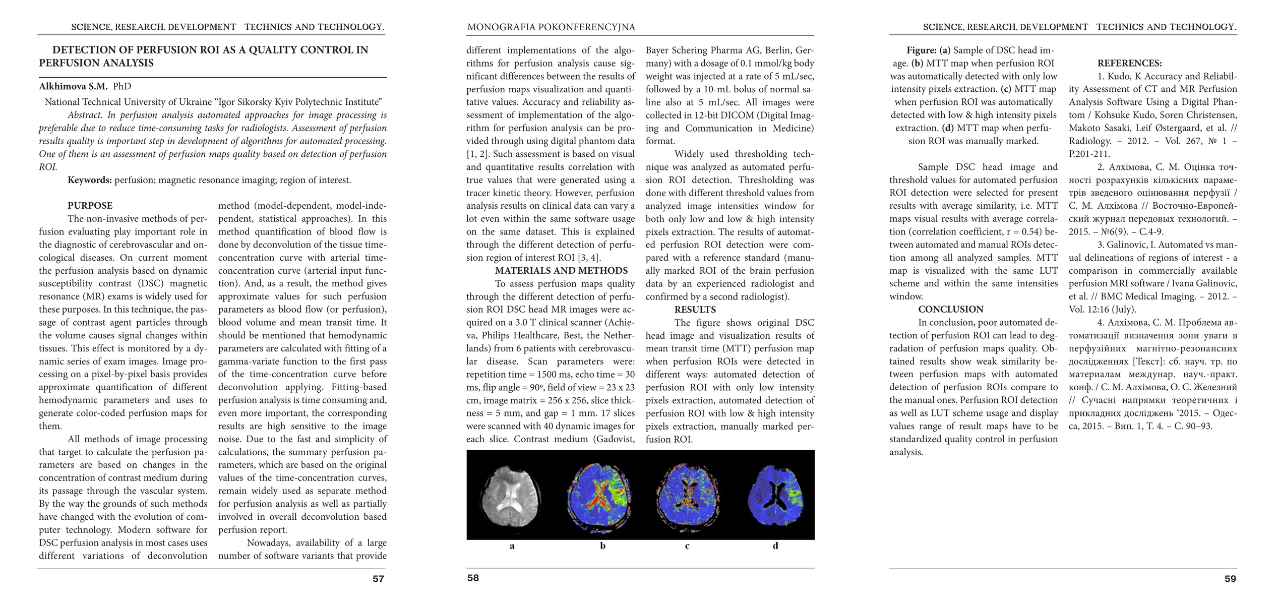
Detection of perfusion ROI as a quality control in perfusion analysis
Publication
Metrics
AI Quick Summary
Summary: This paper discusses automated image processing methods in perfusion analysis to reduce radiologist workload. It emphasizes the importance of assessing perfusion map quality through the detection of perfusion regions of interest (ROI) as a quality control measure in developing automated algorithms.
Paper Preview
Abstract
In perfusion analysis automated approaches for image processing is preferable due to reduce time-consuming tasks for radiologists. Assessment of perfusion results quality is important step in development of algorithms for automated processing. One of them is an assessment of perfusion maps quality based on detection of perfusion ROI.
AI Key Findings
Get AI-generated insights about this paper's methodology, results, significance, and more — seven facets brought into focus.
Impact
Paper Details
PDF Preview
Key Terms
Citation Network
Current paper (gray), citations (green), references (blue)
Display is limited for performance on very large graphs.
Discussion 0