Quick Summary:

This paper demonstrates that nuclear magnetic resonance diffusion experiments can detect the confining boundary of closed pores using modified Stejskal-Tanner magnetic field gradients, which preserve phase information and enhance signal-to-noise ratio, thus providing a method to image the average pore structure in porous media.

Summary

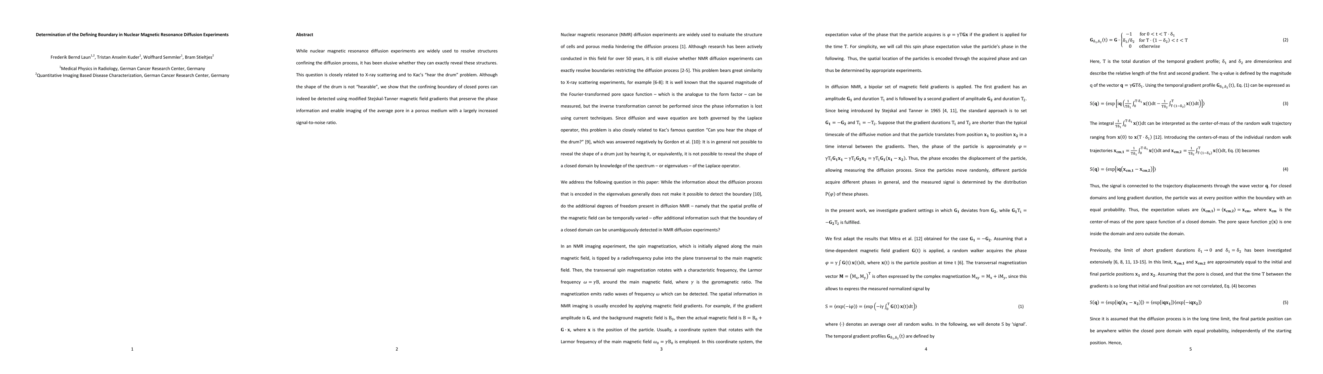
While nuclear magnetic resonance diffusion experiments are widely used to resolve structures confining the diffusion process, it has been elusive whether they can exactly reveal these structures. This question is closely related to X-ray scattering and to Kac's "hear the drum" problem. Although the shape of the drum is not "hearable", we show that the confining boundary of closed pores can indeed be detected using modified Stejskal-Tanner magnetic field gradients that preserve the phase information and enable imaging of the average pore in a porous medium with a largely increased signal-to-noise ratio.

AI Key Findings

Get AI-generated insights about this paper's methodology, results, and significance.

Paper Details

PDF Preview

Download PDF

Key Terms

confining (0.325)
diffusion (0.248)
diffusion (0.230)
structures (0.211)
structures (0.205)
elusive (0.198)
noise ratio (0.191)
closely related (0.190)
magnetic resonance (0.188)
magnetic (0.187)
signal noise (0.185)
preserve (0.184)
magnetic (0.181)
resolve (0.181)
noise ratio (0.180)
preserve (0.180)
closely related (0.177)
signal noise (0.176)
closely (0.174)
largely (0.173)
Title Authors Year Actions
Data inversion of multi-dimensional magnetic resonance in porous media
Fangrong Zong, Huabing Liu, Ruiliang Bai, P. Galvosas
2023
View Paper
Diffusion within pores fully revealed by magnetic resonance.
E. Özarslan, Cem Yolcu, Alfredo Ordinola, Deneb Boito, T. Dela Haije, Mathias Højgaard Jensen, M. Herberthson
2022
View Paper
Flow-compensated diffusion encoding in MRI for improved liver metastasis detection
F. Laun, Tobit Führes, Hannes Seuss, Astrid Müller, S. Bickelhaupt, A. Stemmer, T. Benkert, M. Uder, M. Saake
2022
View Paper
Diffusion pore imaging in the presence of extraporal water.
D. Ludwig, F. Laun, K. Klika, J. Rauch, M. Ladd, P. Bachert, T. Kuder
2021
View Paper
Apparent exchange rate imaging: On its applicability and the connection to the real exchange rate
D. Ludwig, F. Laun, M. Ladd, P. Bachert, T. Kuder
2021
View Paper
MR cell size imaging with temporal diffusion spectroscopy.
Xiaoyu Jiang, Hua Li, Sean P. Devan, J. Gore, Junzhong Xu
2020
View Paper
The sensitivity of diffusion MRI to microstructural properties and experimental factors
M. Afzali, Tomasz Pieciak, Sharlene D. Newman, E. Garyfallidis, E. Özarslan, Hu Cheng, Derek K. Jones
2020
View Paper
Diffusion NMR in periodic media: efficient computation and spectral properties
Nicolas Moutal, Antoine Moutal, D. Grebenkov
2020
View Paper
[Multidimensional diffusion-weighted magnetic resonance imaging].
F. Laun
2019
View Paper
Towards Optimal Sampling in Diffusion MRI
H. Knutsson
2019
View Paper
Stimulated echo double diffusion encoded imaging of closed pores: Influence and removal of unbalanced terms.
Kerstin Demberg, F. Laun, P. Bachert, M. Ladd, T. Kuder
2019
View Paper
Localization regime in diffusion NMR: Theory and experiments.
Nicolas Moutal, Kerstin Demberg, D. Grebenkov, T. Kuder
2019
View Paper
Imaging brain microstructure with diffusion MRI: practicality and applications
D. Alexander, T. Dyrby, M. Nilsson, Hui Zhang
2019
View Paper
Spin echoes: full numerical solution and breakdown of approximative solutions
Christian H. Ziener, T. Kampf, H. Schlemmer, L. Buschle
2019
View Paper
Multimodal integration of diffusion MRI for better characterization of tissue biology
Chunlei Liu, E. Özarslan
2018
View Paper
Axon Diameters and Myelin Content Modulate Microscopic Fractional Anisotropy at Short Diffusion Times in Fixed Rat Spinal Cord
N. Shemesh
2018
View Paper
Experimental determination of pore shapes using phase retrieval from q-space NMR diffraction.
Kerstin Demberg, F. Laun, M. Bertleff, P. Bachert, T. Kuder
2018
View Paper
1D and 2D diffusion pore imaging on a preclinical MR system using adaptive rephasing: Feasibility and pulse sequence comparison.
M. Bertleff, S. Domsch, F. Laun, T. Kuder, L. Schad
2017
View Paper
Microscopic interpretation and generalization of the Bloch-Torrey equation for diffusion magnetic resonance.
Inbar Seroussi, D. Grebenkov, O. Pasternak, N. Sochen
2017
View Paper
Dynamics of local magnetization in the eigenbasis of the Bloch-Torrey operator.
M. Herberthson, E. Özarslan, H. Knutsson, C. Westin
2017
View Paper
Phase incremented echo train acquisition applied to magnetic resonance pore imaging.
Stefan Andreas Hertel, P. Galvosas
2016
View Paper
Quantifying brain microstructure with diffusion MRI: Theory and parameter estimation
D. Novikov, E. Fieremans, S. Jespersen, V. Kiselev
2016
View Paper
Q‐space truncation and sampling in diffusion spectrum imaging
Q. Tian, Ariel S. Rokem, R. Folkerth, A. Nummenmaa, Q. Fan, B. Edlow, J. McNab
2016
View Paper
Homogenized and analytical models for the diffusion MRI signal. (Modélisation du signal de l'IRM de diffusion par des techniques analytiques et d'homogénéisation)
S. Schiavi
2016
View Paper
Observation of structural universality in disordered systems using bulk diffusion measurement.
A. Papaioannou, D. Novikov, E. Fieremans, G. Boutis
2016
View Paper
k and q Dedicated to Paul Callaghan.
B. Blümich
2016
View Paper
Pore sizes and directionality in microcapillaries from angular double-pulsed-field-gradient NMR
D. Morozov, Leah Bar, N. Sochen, Y. Cohen
2016
View Paper
Conventions and nomenclature for double diffusion encoding NMR and MRI
N. Shemesh, S. Jespersen, D. Alexander, Y. Cohen, I. Drobnjak, T. Dyrby, J. Finsterbusch, M. Koch, T. Kuder, F. Laun, M. Lawrenz, H. Lundell, P. Mitra, M. Nilsson, E. Özarslan, D. Topgaard, C. Westin
2016
View Paper
Symmetry of the gradient profile as second experimental dimension in the short-time expansion of the apparent diffusion coefficient as measured with NMR diffusometry.
F. Laun, T. Kuder, Fangrong Zong, S. Hertel, P. Galvosas
2015
View Paper
Microstructural information from angular double‐pulsed‐field‐gradient NMR: From model systems to nerves
D. Morozov, Leah Bar, N. Sochen, Y. Cohen
2015
View Paper
Pulsed second order fields for parallel acquisition of q-space
W. Kittler, P. Galvosas, M. Hunter
2015
View Paper
Magnetic Resonance Pore Imaging: Overcoming the resolution limit of MRI for closed pore systems
S. Hertel, M. Hunter, P. Galvosas
2015
View Paper
Tissue Microstructure Imaging with Diffusion MRI
G. Nedjati-Gilani, D. Alexander
2015
View Paper
Nuclear Magnetic Resonance Upstream Applications: Crude Oil Characterization, Water–Oil Interface Behavior, and Porous Media
T. Lehmann, V. Alvarado
2015
View Paper
Pulsed second order field NMR for real time PGSE and single-shot surface to volume ratio measurements.
W. Kittler, S. Obruchkov, P. Galvosas, M. Hunter
2014
View Paper
Nonparametric pore size distribution using d-PFG: comparison to s-PFG and migration to MRI.
D. Benjamini, M. Komlosh, P. Basser, U. Nevo
2014
View Paper
Parallel acquisition of q-space using second order magnetic fields for single-shot diffusion measurements.
W. Kittler, P. Galvosas, M. W. Hunter, M. W. Hunter
2014
View Paper
Measuring small compartment dimensions by probing diffusion dynamics via Non-uniform Oscillating-Gradient Spin-Echo (NOGSE) NMR.
N. Shemesh, G. Álvarez, L. Frydman
2013
View Paper
Coherent dynamical recoupling of diffusion-driven decoherence in magnetic resonance.
G. Álvarez, N. Shemesh, L. Frydman
2013
View Paper
Magnetic Resonance Pore Imaging, a new tool for porous media research
S. Hertel
2013
View Paper
Diffusion pore imaging by hyperpolarized xenon-129 nuclear magnetic resonance.
T. Kuder, P. Bachert, J. Windschuh, F. Laun
2012
View Paper
NMR-based diffusion pore imaging.
F. Laun, T. Kuder, A. Wetscherek, B. Stieltjes, W. Semmler
2012
View Paper
Diffusion pore imaging with generalized temporal gradient profiles.
F. Laun, T. Kuder
2012
View Paper
Restricted diffusion in NMR in arbitrary inhomogeneous magnetic fields and an application to circular layers.
F. Laun
2012
View Paper
NMR‐based diffusion pore imaging by double wave vector measurements
T. Kuder, F. Laun
2012
View Paper
Measuring Microstructural Features Using Diffusion MRI
N. Shemesh
2020
View Paper
Effective Potential for Magnetic Resonance Measurements of Restricted Diffusion
E. Özarslan, Cem Yolcu, M. Herberthson, C. Westin, H. Knutsson
2017
View Paper
Measuring microscopic anisotropy with diffusion magnetic resonance: From material science to biomedical imaging
A. Ianuş, N. Shemesh, D. Alexander, I. Drobnjak
2017
View Paper
Novel Magnetic Resonance Acquisition and Processing Strategies for Biological Tissue Characterisation
Fangrong Zong
2016
View Paper
From the microstructure to diffusion MRI, and back
D. Grebenkov
2015
View Paper
Introduction to Diffusion Imaging
B. Stieltjes, R. Brunner, K. Fritzsche, F. Laun
2013
View Paper

Comments (0)

This website uses cookies to ensure you get the best experience. Learn more