Development of an algorithm for medical image segmentation of bone tissue in interaction with metallic implants
Publication
Metrics
AI Quick Summary
This study developed an AI-based U-Net algorithm for segmenting bone tissue in contact with metallic implants in microtomography images, achieving 98% accuracy. The method estimates bone growth more accurately than conventional techniques, though it has room for improvement with larger datasets.
Paper Preview
Abstract
This preliminary study focuses on the development of a medical image segmentation algorithm based on artificial intelligence for calculating bone growth in contact with metallic implants. %as a result of the problem of estimating the growth of new bone tissue due to artifacts. %the presence of various types of distortions and errors, known as artifacts. Two databases consisting of computerized microtomography images have been used throughout this work: 100 images for training and 196 images for testing. Both bone and implant tissue were manually segmented in the training data set. The type of network constructed follows the U-Net architecture, a convolutional neural network explicitly used for medical image segmentation. In terms of network accuracy, the model reached around 98\%. Once the prediction was obtained from the new data set (test set), the total number of pixels belonging to bone tissue was calculated. This volume is around 15\% of the volume estimated by conventional techniques, which are usually overestimated. This method has shown its good performance and results, although it has a wide margin for improvement, modifying various parameters of the networks or using larger databases to improve training.
AI Key Findings
Get AI-generated insights about this paper's methodology, results, significance, and more — seven facets brought into focus.
Impact
Paper Details
Authors
PDF Preview
Key Terms
Citation Network
Current paper (gray), citations (green), references (blue)
Display is limited for performance on very large graphs.
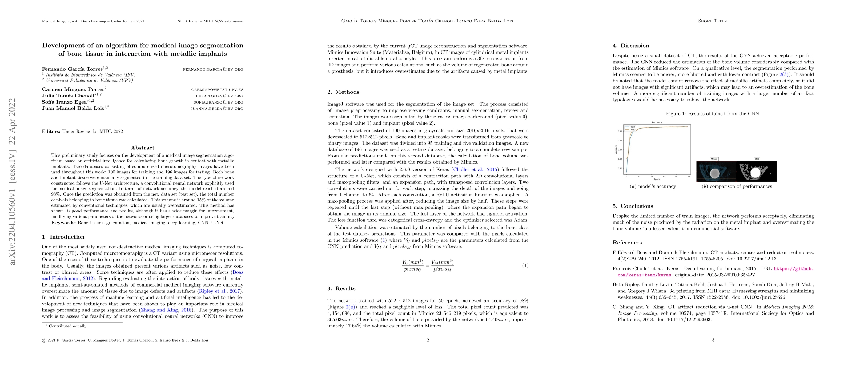
Discussion 0