Quick Summary:

This paper develops an ontology for an integrated image analysis platform to facilitate global sharing of microscopy imaging data. The authors utilize the Resource Description Framework (RDF) to translate the OME data model, enhancing it with 18 upper-level concepts to cover optical and electron microscopy, phenotype data, biosamples, and imaging conditions.

Summary

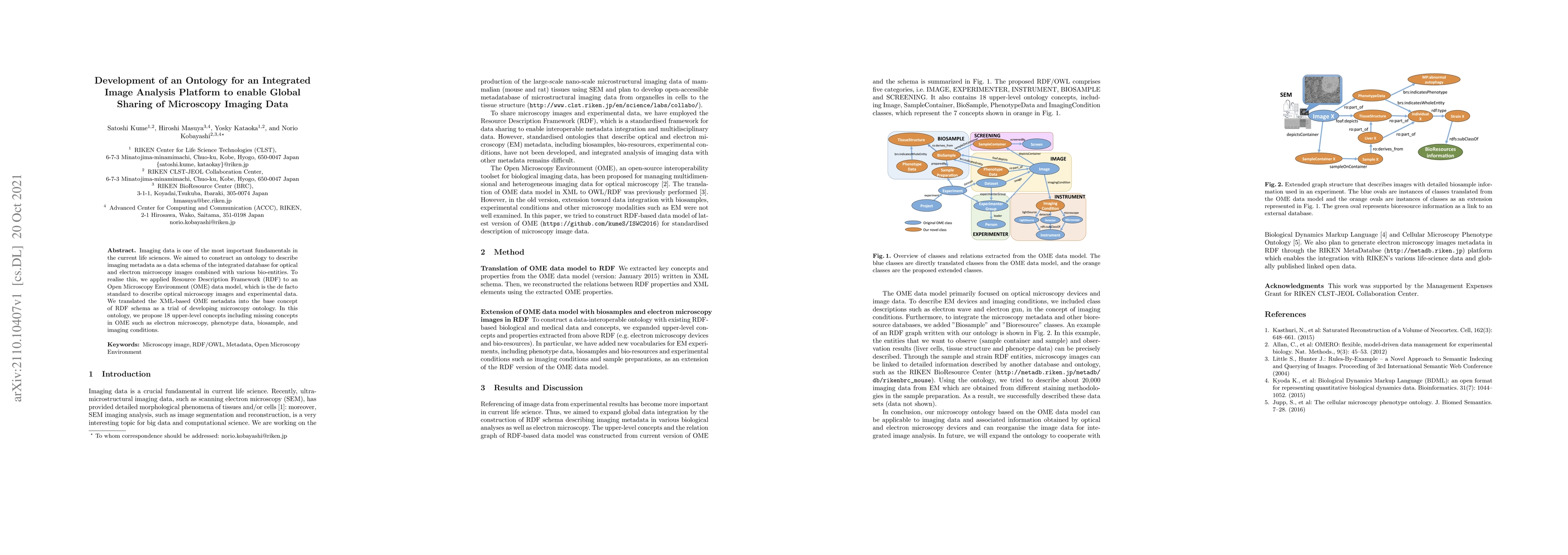
Imaging data is one of the most important fundamentals in the current life sciences. We aimed to construct an ontology to describe imaging metadata as a data schema of the integrated database for optical and electron microscopy images combined with various bio-entities. To realise this, we applied Resource Description Framework (RDF) to an Open Microscopy Environment (OME) data model, which is the de facto standard to describe optical microscopy images and experimental data. We translated the XML-based OME metadata into the base concept of RDF schema as a trial of developing microscopy ontology. In this ontology, we propose 18 upper-level concepts including missing concepts in OME such as electron microscopy, phenotype data, biosample, and imaging conditions.

AI Key Findings

Get AI-generated insights about this paper's methodology, results, and significance.

Paper Details

PDF Preview

Download PDF

Key Terms

microscopy (0.442)
microscopy (0.417)
imaging (0.279)
imaging (0.278)
concepts (0.259)
concepts (0.257)
images (0.205)
images (0.199)
electron (0.198)
electron (0.195)
data (0.189)
optical (0.189)
optical (0.187)
sciences (0.187)
data (0.185)
sciences (0.184)
trial (0.181)
aimed (0.178)
entities (0.176)
entities (0.175)
Title Authors Year Actions
Morphomics via next-generation electron microscopy
Raku Son, Kenji Yamazawa, Akiko Oguchi, Mitsuo Suga, Masaru Tamura, M. Yanagita, Yasuhiro Murakawa, Satoshi Kume
2023
View Paper
End-to-End provenance representation for the understandability and reproducibility of scientific experiments using a semantic approach
Sheeba Samuel, B. König-Ries
2022
View Paper
Towards community-driven metadata standards for light microscopy: tiered specifications extending the OME model
Mathias Hammer, Maximiliaan Huisman, Alessandro Rigano, U. Boehm, J. Chambers, N. Gaudreault, A. North, J. A. Pimentel, D. Sudar, P. Bajcsy, C. Brown, A. Corbett, O. Faklaris, J. Lacoste, A. Laude, Glyn Nelson, R. Nitschke, Farzin Farzam, Carlas S. Smith, D. Grunwald, C. Strambio‐De‐Castillia
2021
View Paper
OME-NGFF: scalable format strategies for interoperable bioimaging data
Josh Moore, Chris Allan, S. Besson, Jean-Marie Burel, Erin E. Diel, David Gault, Kevin Kozlowski, Dominik Lindner, M. Linkert, Trevor Manz, W. Moore, Constantin Pape, C. Tischer, J. Swedlow
2021
View Paper
Development of Semantic Web-Based Imaging Database for Biological Morphome
S. Kume, H. Masuya, M. Maeda, M. Suga, Y. Kataoka, Norio Kobayashi
2017
View Paper
Integrative Data Management for Reproducibility of Microscopy Experiments
Sheeba Samuel
2017
View Paper
OMERO.mde in a use case for microscopy metadata harmonization: Facilitating FAIR principles in practical application with metadata annotation tools
S. Kunis, Sebastian Hänsch, Christian Schmidt, Frances Wong, S. Weidtkamp‐Peters
2021
View Paper
On Bringing Bioimaging Data into the Open (World)
Josh Moore, Norio Kobayashi, S. Kunis, Shuichi Onami, J. Swedlow
2019
View Paper
A provenance-based semantic approach to support understandability, reproducibility, and reuse of scientific experiments
Sheeba Samuel
2019
View Paper
RIKEN MetaDatabase: A Database Platform for Health Care and Life Sciences as a Microcosm of Linked Open Data Cloud
Norio Kobayashi, S. Kume, Kai Lenz, H. Masuya
2018
View Paper

Comments (0)

This website uses cookies to ensure you get the best experience. Learn more