ArxivLens

Quick Summary:

This paper develops an open-source software module for (3D) Slicer to enhance visualization during MR-guided interstitial gynecologic brachytherapy, enabling real-time processing of intra-operative MRI data and multi-stage registration of brachytherapy device models to assist physicians.

Journal Reference: Chen and Egger SpringerPlus 2014, 3:167
arXiv Id: 1404.6116
Comments: 9 pages, 6 figures
Date Published: 2014-04-25
Date Updated: 2014-04-25

Categories:

Key Terms (TF-IDF):

software (0.281)
module (0.236)
tumor (0.187)
death (0.184)
insertion (0.177)
placement (0.176)
research (0.175)
template (0.172)
patient (0.171)
cancer (0.169)
intra (0.163)
patients (0.160)
recurrent (0.160)
disease (0.159)
virtual (0.154)
platform (0.153)
open source (0.151)
enabling (0.148)
cause (0.147)
medical (0.145)
License: http://arxiv.org/licenses/nonexclusive-distrib/1.0/

Summary:

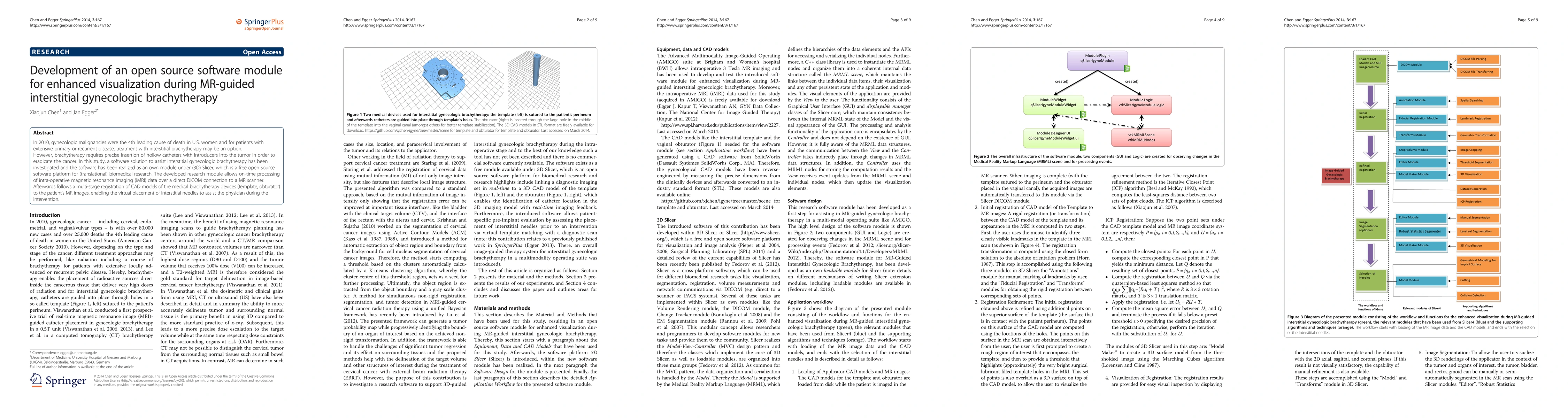
In 2010, gynecologic malignancies were the 4th leading cause of death in U.S. women and for patients with extensive primary or recurrent disease, treatment with interstitial brachytherapy may be an option. However, brachytherapy requires precise insertion of hollow catheters with introducers into the tumor in order to eradicate the cancer. In this study, a software solution to assist interstitial gynecologic brachytherapy has been investigated and the software has been realized as an own module under (3D) Slicer, which is a free open source software platform for (translational) biomedical research. The developed research module allows on-time processing of intra-operative magnetic resonance imaging (iMRI) data over a direct DICOM connection to a MR scanner. Afterwards follows a multi-stage registration of CAD models of the medical brachytherapy devices (template, obturator) to the patient's MR images, enabling the virtual placement of interstitial needles to assist the physician during the intervention.

Citation & Reference Network Visualization

This graph shows how this paper is connected to other research. The current paper is shown in dark gray, papers that cite it are in green, and papers it references are in blue. You can:

Current Paper
Papers citing this work (Citations)
Papers cited by this work (References)

• Hover over any node to see the paper title

• Click a node to view that paper's details

• Drag nodes to rearrange the graph

• Use the zoom controls (bottom right) to zoom in/out

Citations and References

Title Authors Year Actions
Lower jawbone data generation for deep learning tools under MeVisLab
Birgit Pfarrkirchner, Christina Gsaxner, L. Lindner, N. Jakse, J. Wallner, D. Schmalstieg, J. Egger
2018
View Paper
Computer-aided planning and reconstruction of cranial 3D implants
Markus Gall, Xing Li, Xiaojun Chen, D. Schmalstieg, J. Egger
2016
View Paper
Cellular automata segmentation of the boundary between the compacta of vertebral bodies and surrounding structures
J. Egger, C. Nimsky
2016
View Paper
Interactive and scale invariant segmentation of the rectum/sigmoid via user-defined templates
Tobias Lüddemann, J. Egger
2016
View Paper
RFA-cut: Semi-automatic segmentation of radiofrequency ablation zones with and without needles via optimal s-t-cuts
J. Egger, H. Busse, P. Brandmaier, D. Seider, M. Gawlitza, S. Strocka, Philip Voglreiter, Mark Dokter, M. Hofmann, Bernhard Kainz, Xiaojun Chen, A. Hann, Pedro Boechat, Wei Yu, Bernd Freisleben, Tuomas Alhonnoro, M. Pollari, M. Moche, D. Schmalstieg
2015
View Paper
MR diffusion-weighted imaging-based subcutaneous tumour volumetry in a xenografted nude mouse model using 3D Slicer: an accurate and repeatable method
Zelan Ma, Xin Chen, Yanqi Huang, Lan He, Cuishan Liang, C. Liang, Zaiyi Liu
2015
View Paper
Development of a surgical navigation system based on augmented reality using an optical see-through head-mounted display
Xiaojun Chen, Lu Xu, Yiping Wang, Huixiang Wang, Fang Wang, Xiangsen Zeng, Qiugeng Wang, J. Egger
2015
View Paper
Computer-aided planning and reconstruction of cranial 3D implants.
Markus Gall, Xing Li, Xiaojun Chen, D. Schmalstieg, J. Egger
2016
View Paper

Comments (0)

This website uses cookies to ensure you get the best experience. Learn more