Publication
Metrics
AI Quick Summary
The research introduces DFENet, a novel network for accurate brain MRI segmentation to aid stroke diagnosis. DFENet combines 2D and 3D CNN features, uses a parallel partial decoder for contextual information, and an edge-guidance loss for improved learning. Evaluation on the ATLAS dataset shows superior performance compared to existing methods, making it a robust tool for biomedical applications.
Paper Preview
Abstract
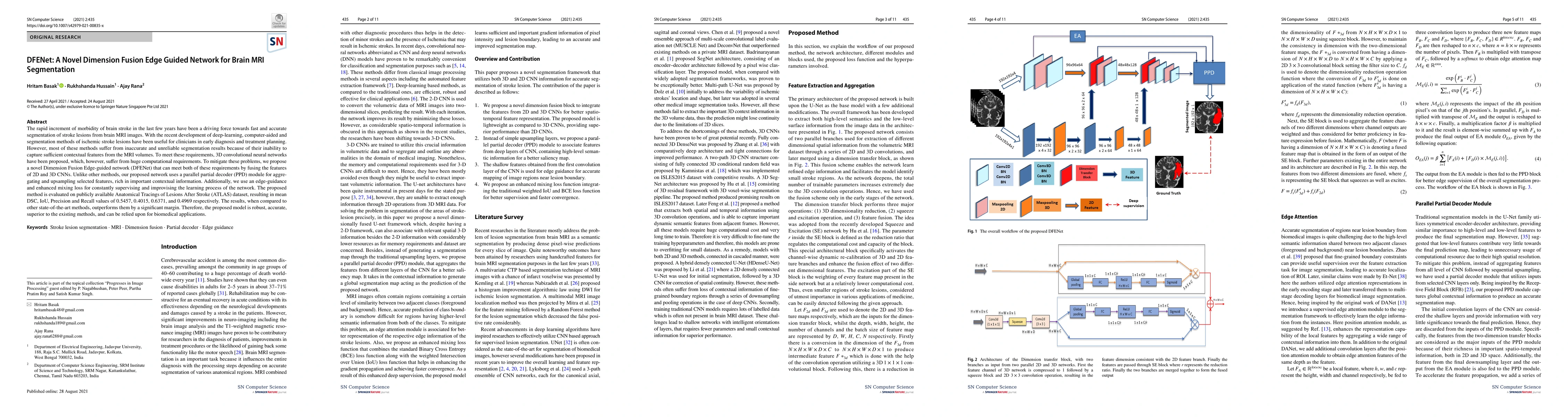
The rapid increment of morbidity of brain stroke in the last few years have been a driving force towards fast and accurate segmentation of stroke lesions from brain MRI images. With the recent development of deep-learning, computer-aided and segmentation methods of ischemic stroke lesions have been useful for clinicians in early diagnosis and treatment planning. However, most of these methods suffer from inaccurate and unreliable segmentation results because of their inability to capture sufficient contextual features from the MRI volumes. To meet these requirements, 3D convolutional neural networks have been proposed, which, however, suffer from huge computational requirements. To mitigate these problems, we propose a novel Dimension Fusion Edge-guided network (DFENet) that can meet both of these requirements by fusing the features of 2D and 3D CNNs. Unlike other methods, our proposed network uses a parallel partial decoder (PPD) module for aggregating and upsampling selected features, rich in important contextual information. Additionally, we use an edge-guidance and enhanced mixing loss for constantly supervising and improvising the learning process of the network. The proposed method is evaluated on publicly available Anatomical Tracings of Lesions After Stroke (ATLAS) dataset, resulting in mean DSC, IoU, Precision and Recall values of 0.5457, 0.4015, 0.6371, and 0.4969 respectively. The results, when compared to other state-of-the-art methods, outperforms them by a significant margin. Therefore, the proposed model is robust, accurate, superior to the existing methods, and can be relied upon for biomedical applications.
AI Key Findings
Get AI-generated insights about this paper's methodology, results, significance, and more — seven facets brought into focus.
Impact
Paper Details
PDF Preview
Key Terms
Citation Network
Current paper (gray), citations (green), references (blue)
Display is limited for performance on very large graphs.
Discussion 0