This research develops an autoencoder-based method to detect Helicobacter pylori in immunohistochemically stained whole slide images using minimal annotations. The method shows high accuracy, sensitivity, and specificity, proving effective even with limited training data, and thus can assist pathologists in quickly identifying infected patches.
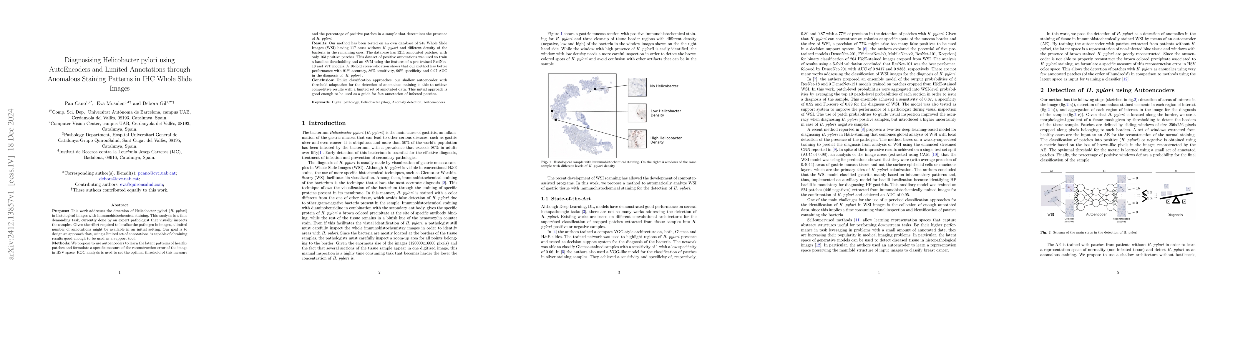
Purpose: This work addresses the detection of Helicobacter pylori (H. pylori) in histological images with immunohistochemical staining. This analysis is a time demanding task, currently done by an expert pathologist that visually inspects the samples. Given the effort required to localise the pathogen in images, a limited number of annotations might be available in an initial setting. Our goal is to design an approach that, using a limited set of annotations, is capable of obtaining results good enough to be used as a support tool. Methods: We propose to use autoencoders to learn the latent patterns of healthy patches and formulate a specific measure of the reconstruction error of the image in HSV space. ROC analysis is used to set the optimal threshold of this measure and the percentage of positive patches in a sample that determines the presence of H. pylori. Results: Our method has been tested on an own database of 245 Whole Slide Images (WSI) having 117 cases without H. pylori and different density of the bacteria in the remaining ones. The database has 1211 annotated patches, with only 163 positive patches. This dataset of positive annotations was used to train a baseline thresholding and an SVM using the features of a pre-trained RedNet18 and ViT models. A 10-fold cross-validation shows that our method has better performance with 91% accuracy, 86% sensitivity, 96% specificity and 0.97 AUC in the diagnosis of H. pylori. Conclusion: Unlike classification approaches, our shallow autoencoder with threshold adaptation for the detection of anomalous staining is able to achieve competitive results with a limited set of annotated data. This initial approach is good enough to be used as a guide for fast annotation of infected patches.
Generated Sep 03, 2025
The research proposes using autoencoders to detect Helicobacter pylori in immunohistochemical (IHC) stained whole slide images (WSIs) by learning normal tissue patterns from non-infected samples. Anomalous staining patterns, particularly the brown color associated with H. pylori, are poorly reconstructed, allowing for anomaly detection. Patches are defined using a sliding window approach, and a metric based on the loss of brown-like pixels in the reconstructed images is used for classification.
This research presents a deep learning (DL) system for H. pylori diagnosis in IHC-stained gastric mucosa, utilizing autoencoders for anomaly detection without requiring extensive annotation. The approach significantly reduces manual cost and time, offering a competitive solution with a limited number of annotated patches.
The research introduces a self-learning anomaly detection approach using autoencoders for H. pylori staining anomalies, requiring minimal annotated patches and providing competitive diagnostic metrics.
This work stands out by employing autoencoders for anomaly detection in a limited annotation setting, offering a more efficient and cost-effective alternative to existing deep learning methods for H. pylori diagnosis in IHC-stained gastric mucosa.
Current paper (gray), citations (green), references (blue)
Display is limited for performance on very large graphs.
Pau Cano, Álvaro Caravaca, Debora Gil et al.
Chen, Zhe, Cui, Lei, Zhang, Lihua et al.
Amit Das, Saeed Hassanpour, Bing Ren et al.
Liu, Xiaoying, Ren, Bing, Ma, Weijie et al.
No citations found for this paper.
Comments (0)