Dictionary-based Method for Vascular Segmentation for OCTA Images
Publication
Metrics
AI Quick Summary
This paper proposes a dictionary-based machine learning method for segmenting larger vessels, capillaries, and background in OCTA images, enabling robust quantification of retinal blood flow. The method is trained on specific parameters using training data and demonstrates effective segmentation of the microvasculature in the retina.
Paper Preview
Abstract
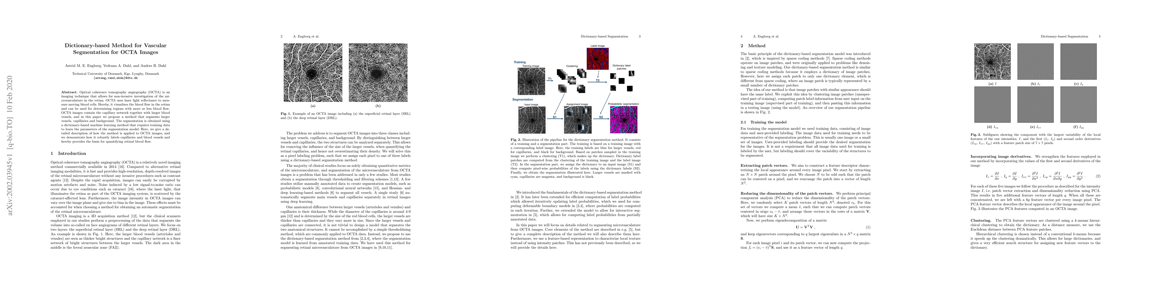
Optical coherence tomography angiography (OCTA) is an imaging technique that allows for non-invasive investigation of the microvasculature in the retina. OCTA uses laser light reflectance to measure moving blood cells. Hereby, it visualizes the blood flow in the retina and can be used for determining regions with more or less blood flow. OCTA images contain the capillary network together with larger blood vessels, and in this paper we propose a method that segments larger vessels, capillaries and background. The segmentation is obtained using a dictionary-based machine learning method that requires training data to learn the parameters of the segmentation model. Here, we give a detailed description of how the method is applied to OCTA images, and we demonstrate how it robustly labels capillaries and blood vessels and hereby provides the basis for quantifying retinal blood flow.
AI Key Findings
Get AI-generated insights about this paper's methodology, results, significance, and more — seven facets brought into focus.
Impact
Paper Details
Authors
PDF Preview
Key Terms
Citation Network
Current paper (gray), citations (green), references (blue)
Display is limited for performance on very large graphs.
Discussion 0