Diff-CL: A Novel Cross Pseudo-Supervision Method for Semi-supervised Medical Image Segmentation

Source: ArXiv

AI Quick Summary

Diff-CL introduces a novel cross pseudo-supervision method combining diffusion models and convolutional networks for semi-supervised medical image segmentation, leveraging distributional and detailed information to achieve robust and accurate results, achieving state-of-the-art performance across multiple datasets.

Share: Share on X LinkedIn

Summary

Semi-supervised learning utilizes insights from unlabeled data to improve model generalization, thereby reducing reliance on large labeled datasets. Most existing studies focus on limited samples and fail to capture the overall data distribution. We contend that combining distributional information with detailed information is crucial for achieving more robust and accurate segmentation results. On the one hand, with its robust generative capabilities, diffusion models (DM) learn data distribution effectively. However, it struggles with fine detail capture, leading to generated images with misleading details. Combining DM with convolutional neural networks (CNNs) enables the former to learn data distribution while the latter corrects fine details. While capturing complete high-frequency details by CNNs requires substantial computational resources and is susceptible to local noise. On the other hand, given that both labeled and unlabeled data come from the same distribution, we believe that regions in unlabeled data similar to overall class semantics to labeled data are likely to belong to the same class, while regions with minimal similarity are less likely to. This work introduces a semi-supervised medical 
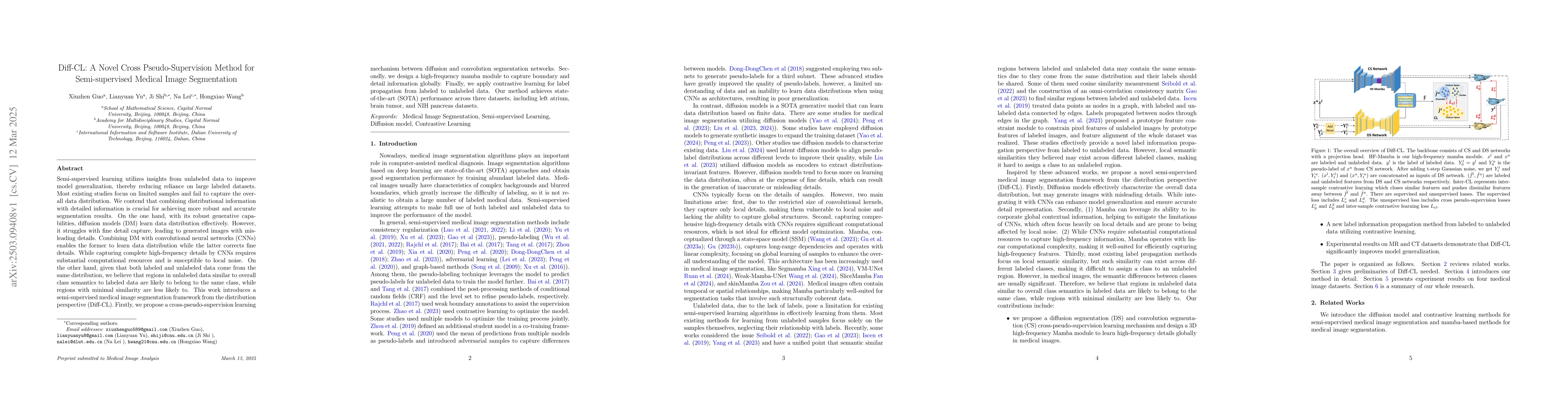
image segmentation framework from the distribution perspective (Diff-CL). Firstly, we propose a cross-pseudo-supervision learning mechanism between diffusion and convolution segmentation networks. Secondly, we design a high-frequency mamba module to capture boundary and detail information globally. Finally, we apply contrastive learning for label propagation from labeled to unlabeled data. Our method achieves state-of-the-art (SOTA) performance across three datasets, including left atrium, brain tumor, and NIH pancreas datasets.

AI Key Findings

Generated Jun 10, 2025

Methodology

The research proposes a semi-supervised medical image segmentation framework (Diff-CL) from a distribution perspective, combining diffusion models (DM) with convolutional neural networks (CNNs) and using contrastive learning for label propagation.

Key Results

  • Diff-CL achieves state-of-the-art performance across three datasets: left atrium, brain tumor, and NIH pancreas.
  • Diff-CL outperforms nine other semi-supervised image segmentation methods on left atrium (LA) and brain tumor (BraTS) datasets under various labeled data settings.
  • Diff-CL significantly improves segmentation accuracy and overlap (Dice and Jaccard) on pancreas dataset compared to baseline methods.

Significance

This research is significant as it addresses the limitations of existing semi-supervised methods by effectively combining distributional and detailed information, reducing reliance on large labeled datasets, and improving segmentation accuracy in medical imaging.

Technical Contribution

The introduction of a cross-pseudo-supervision learning mechanism between diffusion and convolution segmentation networks, a high-frequency mamba module for detail learning, and contrastive learning for label propagation.

Novelty

Diff-CL stands out by integrating diffusion models with CNNs for distribution and detail learning, respectively, and using contrastive learning for label propagation, which hasn't been extensively explored in semi-supervised medical image segmentation.

Limitations

  • The method requires substantial computational resources for capturing high-frequency details using CNNs.
  • While Diff-CL performs well in accuracy prediction, it needs more attention to boundary precision, especially on the pancreas dataset.

Future Work

  • Explore more efficient methods to capture high-frequency details with reduced computational costs.
  • Investigate strategies to improve boundary precision and contour smoothness in segmentation results.

Paper Details

PDF Preview

Download PDF

Citation Network

Current paper (gray), citations (green), references (blue)

Display is limited for performance on very large graphs.

Current Paper
Citations
References
Click to view

No citations found for this paper.

Comments (0)

This website uses cookies to ensure you get the best experience. Learn more