Summary
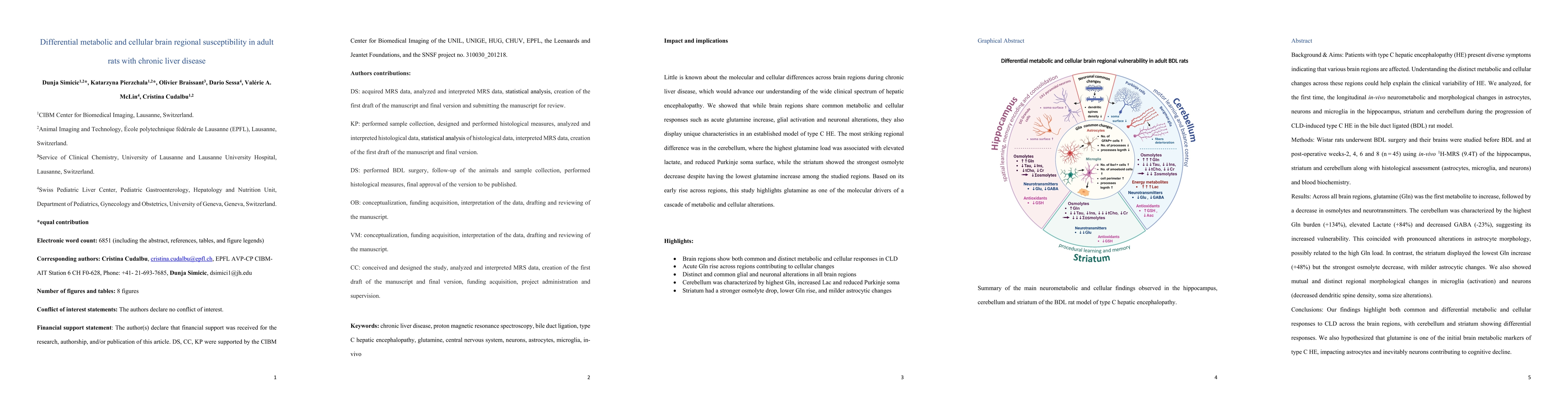
Background & Aims: Patients with type C hepatic encephalopathy (HE) present diverse symptoms indicating that various brain regions are affected. Understanding the distinct metabolic and cellular changes across these regions could help explain the clinical variability of HE. We analyzed, for the first time, the longitudinal in-vivo neurometabolic and morphological changes in astrocytes, neurons and microglia in the hippocampus, striatum and cerebellum during the progression of CLD-induced type C HE in the bile duct ligated (BDL) rat model. Methods: Wistar rats underwent BDL and their brains were studied before BDL and at post-operative weeks-2, 4, 6 and 8 using in-vivo 1H-MRS (9.4T) of the hippocampus, striatum and cerebellum along with histological assessment (astrocytes, microglia, and neurons) and blood biochemistry. Results: Across all brain regions, glutamine (Gln) was the first metabolite to increase, followed by a decrease in osmolytes and neurotransmitters. The cerebellum was characterized by the highest Gln burden (+134%), elevated Lactate (+84%) and decreased GABA (-23%), suggesting its increased vulnerability. This coincided with pronounced alterations in astrocyte morphology, possibly related to the high Gln load. In contrast, the striatum displayed the lowest Gln increase (+48%) but the strongest osmolyte decrease, with milder astrocytic changes. We also showed mutual and distinct regional morphological changes in microglia (activation) and neurons (decreased dendritic spine density, soma size alterations). Conclusions: Our findings highlight both common and differential metabolic and cellular responses to CLD across the brain regions, with cerebellum and striatum showing differential responses. We also hypothesized that glutamine is one of the initial brain metabolic markers of type C HE, impacting astrocytes and inevitably neurons contributing to cognitive decline.
AI Key Findings
Generated Jun 10, 2025
Methodology
The study utilized a bile duct ligation (BDL) rat model of chronic liver disease (CLD)-induced type C hepatic encephalopathy (HE), combining longitudinal in-vivo 1H-MRS and ex-vivo analysis of brain cell subtypes in Wistar rats. The research tracked metabolic and cellular responses in the cerebellum, hippocampus, and striatum over 8 weeks post-BDL.
Key Results
- Glutamine (Gln) was identified as the first globally increased metabolite in all three brain regions, suggesting its role as a surrogate marker for the disease.
- The cerebellum exhibited the heaviest Gln burden, elevated lactate, decreased GABA, and pronounced astrocytic and microglial changes, indicating increased vulnerability.
- The striatum displayed the lowest Gln increase but the strongest decrease in osmolytes, with milder astrocytic changes and distinct microglial and neuronal alterations.
- Neuronal morphological changes were observed, including decreased dendritic spine density and soma size alterations in hippocampal CA1 and DG neurons, as well as cerebellar Purkinje cells.
- Astrocytic reactivity, osmotic stress, and oxidative stress were identified as significant factors in the progression of HE.
Significance
This research highlights the differential metabolic and cellular responses to CLD across brain regions, emphasizing the cerebellum and striatum's differential responses. It suggests that glutamine is an initial brain metabolic marker of type C HE, impacting astrocytes and neurons, which may contribute to cognitive decline.
Technical Contribution
The study presents a comprehensive analysis of in-vivo neurometabolic and morphological changes in astrocytes, neurons, and microglia across different brain regions in a rat model of CLD-induced type C HE.
Novelty
This research is novel in its longitudinal in-vivo 1H-MRS analysis combined with histological assessment, providing insights into the distinct metabolic and cellular changes in various brain regions during the progression of CLD-induced type C HE.
Limitations
- The study was conducted on a rat model, which may not fully replicate human HE.
- Longitudinal 1H-MRS measurements were limited to three brain regions, potentially overlooking other affected areas.
Future Work
- Further investigation into the role of glutamine as an early marker for HE and its impact on neuronal function.
- Exploration of therapeutic interventions targeting astrocytic and neuronal alterations to mitigate cognitive decline in HE.
Paper Details
PDF Preview
Similar Papers
Found 4 papersLipid Accumulation and Insulin Resistance: Bridging Metabolic Dysfunction-Associated Fatty Liver Disease and Chronic Kidney Disease.
Wang, Na, Yang, Min, Cao, Xinyi et al.
Alistipes putredinis Ameliorates Metabolic Dysfunction-Associated Steatotic Liver Disease in Rats via Gut Microbiota Remodeling and Inflammatory Suppression.
Yang, Jing, Lu, Yao, Xu, Jianguo et al.
No citations found for this paper.
Comments (0)