Diffraction imaging for in-situ characterization of double-crystal x-ray monochromators
Publication
Metrics
AI Quick Summary
This paper proposes and validates an in-situ diffraction imaging method for characterizing double-crystal x-ray monochromators under synchrotron radiation, providing rapid evaluation of the exit beam wavefront and local misorientation of crystal planes. This method complements finite element analysis and offers insights into intrinsic crystal quality.
Paper Preview
Abstract
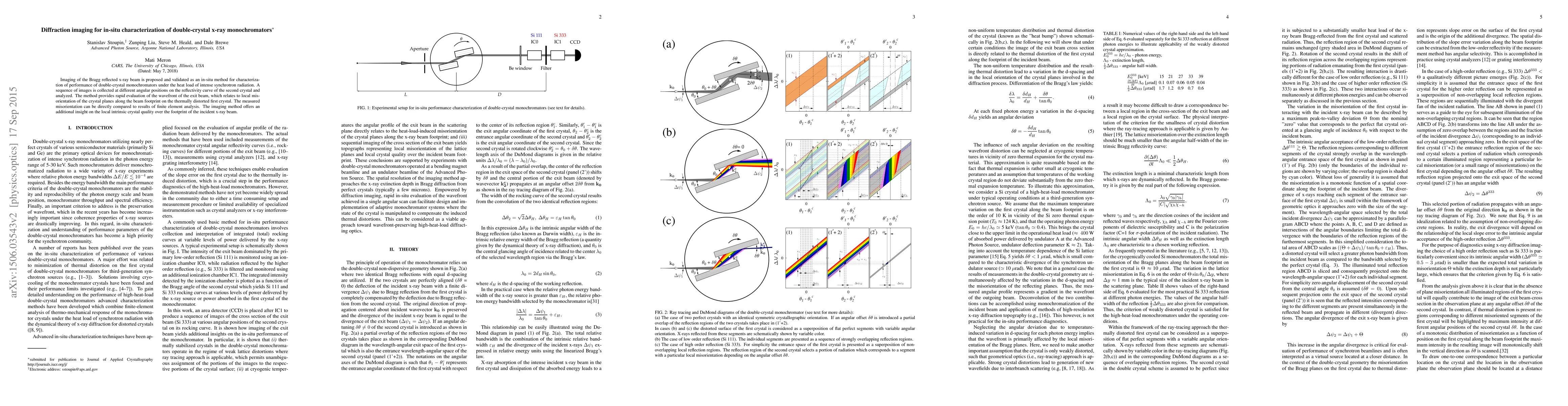
Imaging of the Bragg reflected x-ray beam is proposed and validated as an in-situ method for characterization of performance of double-crystal monochromators under the heat load of intense synchrotron radiation. A sequence of images is collected at different angular positions on the reflectivity curve of the second crystal and analyzed. The method provides rapid evaluation of the wavefront of the exit beam, which relates to local misorientation of the crystal planes along the beam footprint on the thermally distorted first crystal. The measured misorientation can be directly compared to results of finite element analysis. The imaging method offers an additional insight on the local intrinsic crystal quality over the footprint of the incident x-ray beam.
AI Key Findings
Get AI-generated insights about this paper's methodology, results, significance, and more — seven facets brought into focus.
Impact
Paper Details
PDF Preview
Key Terms
Citation Network
Current paper (gray), citations (green), references (blue)
Display is limited for performance on very large graphs.
Discussion 0