Diffraction tomography with Fourier ptychography
Publication
Metrics
Paper Preview
Abstract
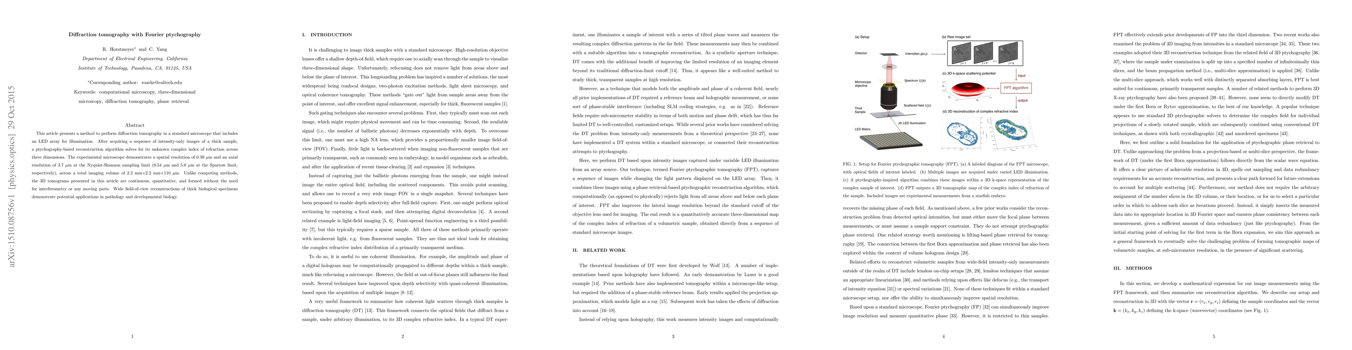
This article presents a method to perform diffraction tomography in a standard microscope that includes an LED array for illumination. After acquiring a sequence of intensity-only images of a thick sample, a ptychography-based reconstruction algorithm solves for its unknown complex index of refraction across three dimensions. The experimental microscope demonstrates a spatial resolution of 0.39 $\mu$m and an axial resolution of 3.7 $\mu$m at the Nyquist-Shannon sampling limit (0.54 $\mu$m and 5.0 $\mu$m at the Sparrow limit, respectively), across a total imaging volume of 2.2 mm $\times$ 2.2 mm $\times$ 110 $\mu$m. Unlike competing methods, the 3D tomograms presented in this article are continuous, quantitative, and formed without the need for interferometry or any moving parts. Wide field-of-view reconstructions of thick biological specimens demonstrate potential applications in pathology and developmental biology.
AI Key Findings
Get AI-generated insights about this paper's methodology, results, significance, and more — seven facets brought into focus.
Impact
Paper Details
PDF Preview
Key Terms
Citation Network
Current paper (gray), citations (green), references (blue)
Display is limited for performance on very large graphs.
Discussion 0