ArxivLens

Quick Summary:

This paper presents a low-cost, portable diffuser-based computational funduscope that reconstructs clinical features of a model eye with shift-invariance and invariant magnification. The system demonstrates fundus image reconstruction over a 33° field-of-view and robustness to refractive errors, potentially enabling combined ocular aberrometry and funduscopic screening.

Journal Reference: Opt. Express 28, 19641-19654 (2020)
DOI: 10.1364/OE.395112
arXiv Id: 2002.11911
Date Published: 2020-06-23
Date Updated: 2020-06-23

Key Terms (TF-IDF):

enable (0.207)
combined (0.192)
image (0.168)
refractive (0.165)
circ (0.158)
low cost (0.154)
reconstruct (0.152)
care (0.152)
features (0.152)
diagnostic (0.151)
lens (0.150)
compared existing (0.149)
screening (0.149)
conjugate (0.147)
clinical (0.144)
poor (0.143)
range (0.139)
sensor (0.139)
spread (0.135)
focused (0.133)
License: http://arxiv.org/licenses/nonexclusive-distrib/1.0/

Summary:

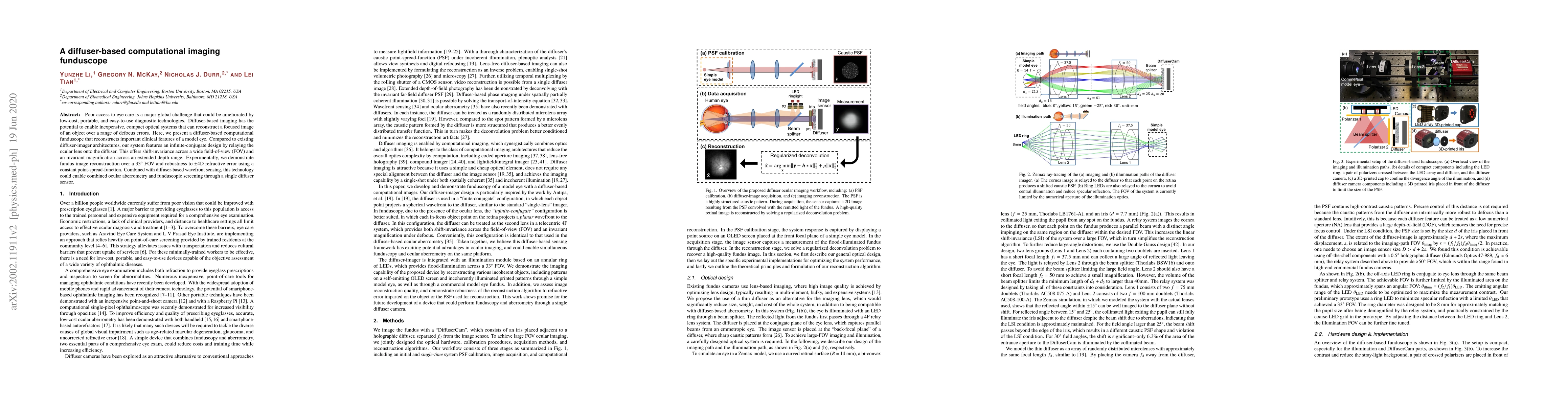
Poor access to eye care is a major global challenge that could be ameliorated by low-cost, portable, and easy-to-use diagnostic technologies. Diffuser-based imaging has the potential to enable inexpensive, compact optical systems that can reconstruct a focused image of an object over a range of defocus errors. Here, we present a diffuser-based computational funduscope that reconstructs important clinical features of a model eye. Compared to existing diffuser-imager architectures, our system features an infinite-conjugate design by relaying the ocular lens onto the diffuser. This offers shift-invariance across a wide field-of-view (FOV) and an invariant magnification across an extended depth range. Experimentally, we demonstrate fundus image reconstruction over a 33$^{\circ}$ FOV and robustness to $\pm$4D refractive error using a constant point-spread-function. Combined with diffuser-based wavefront sensing, this technology could enable combined ocular aberrometry and funduscopic screening through a single diffuser sensor.

Citation & Reference Network Visualization

This graph shows how this paper is connected to other research. The current paper is shown in dark gray, papers that cite it are in green, and papers it references are in blue. You can:

Current Paper
Papers citing this work (Citations)
Papers cited by this work (References)

• Hover over any node to see the paper title

• Click a node to view that paper's details

• Drag nodes to rearrange the graph

• Use the zoom controls (bottom right) to zoom in/out

Citations and References

Title Authors Year Actions
Advances in lensless imaging based on machine learning using ray tracing
Samira Arabpou, Simon Thibault
2025
View Paper
Ophthalmic fundus camera design based on freeform surface for reducing refractive error sensitivity
Wenchao Zhang, Weilin Chen, Jun Chang, Yi Huang, Xuehui Zhao, Xu-yang Li
2023
View Paper
Different Channels to Transmit Information in a Scattering Medium
Xuyu Zhang, Jingjing Gao, Yu H. Gan, C. Song, Dawei Zhang, S. Zhuang, Shensheng Han, Puxiang Lai, Honglin Liu
2022
View Paper
Smartphone-based imaging systems for medical applications: a critical review
Brady Hunt, Alberto J. Ruiz, B. Pogue
2021
View Paper
Three-dimensional phase optical transfer function in axially symmetric microscopic quantitative phase imaging.
Jianhui Huang, Yijun Bao, T. Gaylord
2020
View Paper
Four-frame pixel super-resolution method for lensless imaging systems
Zhengyu Wu, Gang Chen, Shutian Liu, Wei Liu, Dazhao Chi, Bin Gao, Yutong Li, Zhengjun Liu
View Paper

Similar Papers

In vivo fundus imaging and computational refocusing with a diffuser-based fundus camera

Corey Simmerer, Marisa Morakis, Lei Tian, Lia Gomez-Perez, T. Y. Alvin Liu, Nicholas J. Durr

Published: 2024-06-12

DaISy: Diffuser-aided Sub-THz Imaging System

Ke Chen, Yiyao Zhang, Shao-Hsuan Wu, Shang Hua Yang

Published: 2024-03-07

Comments (0)

This website uses cookies to ensure you get the best experience. Learn more