ArxivLens

Quick Summary:

This study uses free gradient waveforms in diffusion MRI to investigate restricted diffusion and exchange in the human brain, revealing distinct time-dependence signatures in grey and white matter. The findings suggest that grey matter exhibits both restricted diffusion and exchange, while white matter primarily shows restricted diffusion, with exchange being at least twice as fast in grey matter.

arXiv Id: 2304.02764
Date Published: 2023-04-07
Date Updated: 2023-04-07

Categories:

Key Terms (TF-IDF):

exchange (0.298)
exchange (0.295)
waveforms (0.292)
restricted (0.270)
diffusion (0.266)
restricted (0.264)
diffusion (0.258)
signatures (0.246)
signatures (0.243)
white (0.223)
gradient (0.209)
white (0.206)
gradient (0.189)
brain (0.183)
brain (0.182)
matter (0.177)
matter (0.170)
protocol (0.165)
dependence (0.163)
protocol (0.156)
License: http://creativecommons.org/licenses/by/4.0/

Summary:

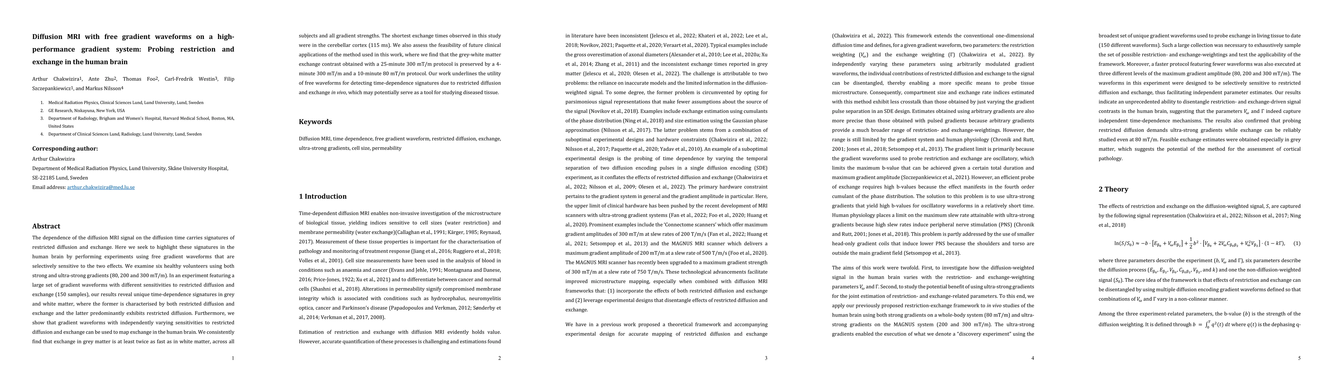
The dependence of the diffusion MRI signal on the diffusion time carries signatures of restricted diffusion and exchange. Here we seek to highlight these signatures in the human brain by performing experiments using free gradient waveforms that are selectively sensitive to the two effects. We examine six healthy volunteers using both strong and ultra-strong gradients (80, 200 and 300 mT/m). In an experiment featuring a large set of gradient waveforms with different sensitivities to restricted diffusion and exchange (150 samples), our results reveal unique time-dependence signatures in grey and white matter, where the former is characterised by both restricted diffusion and exchange and the latter predominantly exhibits restricted diffusion. Furthermore, we show that gradient waveforms with independently varying sensitivities to restricted diffusion and exchange can be used to map exchange in the human brain. We consistently find that exchange in grey matter is at least twice as fast as in white matter, across all subjects and all gradient strengths. The shortest exchange times observed in this study were in the cerebellar cortex (115 ms). We also assess the feasibility of future clinical applications of the method used in this work, where we find that the grey-white matter exchange contrast obtained with a 25-minute 300 mT/m protocol is preserved by a 4-minute 300 mT/m and a 10-minute 80 mT/m protocol. Our work underlines the utility of free waveforms for detecting time-dependence signatures due to restricted diffusion and exchange in vivo, which may potentially serve as a tool for studying diseased tissue.

Citation & Reference Network Visualization

This graph shows how this paper is connected to other research. The current paper is shown in dark gray, papers that cite it are in green, and papers it references are in blue. You can:

Current Paper
Papers citing this work (Citations)
Papers cited by this work (References)

• Hover over any node to see the paper title

• Click a node to view that paper's details

• Drag nodes to rearrange the graph

• Use the zoom controls (bottom right) to zoom in/out

Citations and References

Title Authors Year Actions
In vivo rat brain mapping of multiple gray matter water populations using nonparametric D(ω)‐R 1‐R 2 distributions MRI
Maxime Yon, Omar Narvaez, Daniel Topgaard, Alejandra Sierra
2024
View Paper
Diffusional kurtosis time dependence and the water exchange rate for the multi‐compartment Kärger model
Jens H Jensen
2023
View Paper
Probing brain tissue microstructure with MRI: principles, challenges, and the role of multidimensional diffusion-relaxation encoding
Björn Lampinen, Filip Szczepankiewicz, J. Lätt, Linda Knutsson, J. Mårtensson, I. Björkman-Burtscher, D. Westen, P. Sundgren, F. Ståhlberg, M. Nilsson
2023
View Paper
Quantifying human gray matter microstructure using neurite exchange imaging (NEXI) and 300 mT/m gradients
Quentin Uhl, Tommaso Pavan, Malwina Molendowska, Derek K. Jones, M. Palombo, Ileana Jelescu Department of Radiology, Lausanne University Hospital, Lausanne, Switzerland., School of Biology, Medicine, University of Lausanne, Cardiff University Brain Research Imaging Centre, C. University, Cardiff, United Kingdom., School of Materials Science, Informatics, Uk, joint last authorship
2023
View Paper
Diffusional exchange versus microscopic kurtosis from CTI: Two conflicting interpretations of the same data
Arthur Chakwizira, Filip Szczepankiewicz, Markus Nilsson
2023
View Paper
Can the filter exchange imaging pulse sequence be adapted for applications with thin slices and restricted diffusion?
Samo Lasi ˇ c, Arthur Chakwizira, Henrik Lundell, C. Westin, Markus Nilsson
View Paper

Similar Papers

The role of dendritic spines in water exchange measurements with diffusion MRI: Double Diffusion Encoding and free-waveform MRI

Marco Palombo, Arthur Chakwizira, Filip Szczepankiewicz, Markus Nilsson, Kadir Şimşek

Published: 2025-04-30

Diff5T: Benchmarking Human Brain Diffusion MRI with an Extensive 5.0 Tesla K-Space and Spatial Dataset

Jian Cheng, Shanshan Wang, Qi Yang, Fan Zhang, Hua Guo, Qiyuan Tian, Sen Jia, Hairong Zheng, Yaowen Xing, Shoujun Yu, Changjun Tie, Jiayu Zhu, Haohao Peng, Yijing Dong, Jianzhong He, Xiuqin Jia, Guobin Li

Published: 2024-12-09

Comments (0)

This website uses cookies to ensure you get the best experience. Learn more