This paper presents a novel digital confocal microscopy technique using multimode fibers to enhance imaging deep inside biological tissues. By digitally engineering the excitation wavefront and applying a virtual digital pinhole, the method achieves significantly increased contrast, with a lateral resolution of 1.5 µm and an axial resolution of 12.7 µm.
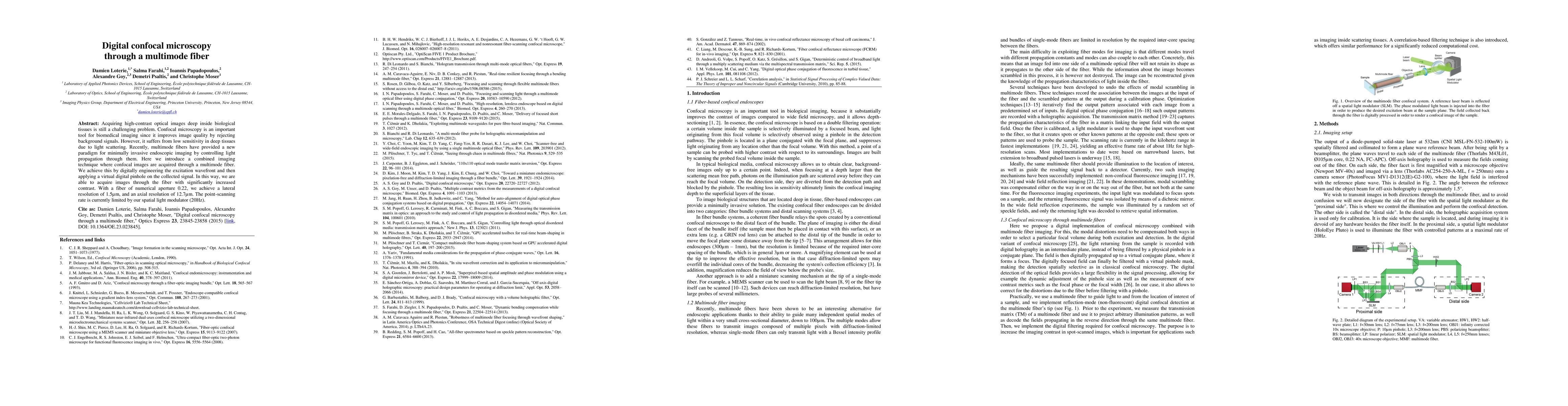
Acquiring high-contrast optical images deep inside biological tissues is still a challenging problem. Confocal microscopy is an important tool for biomedical imaging since it improves image quality by rejecting background signals. However, it suffers from low sensitivity in deep tissues due to light scattering. Recently, multimode fibers have provided a new paradigm for minimally invasive endoscopic imaging by controlling light propagation through them. Here we introduce a combined imaging technique where confocal images are acquired through a multimode fiber. We achieve this by digitally engineering the excitation wavefront and then applying a virtual digital pinhole on the collected signal. In this way, we are able to acquire images through the fiber with significantly increased contrast. With a fiber of numerical aperture 0.22, we achieve a lateral resolution of 1.5um, and an axial resolution of 12.7um. The point-scanning rate is currently limited by our spatial light modulator (20Hz).
Generated Sep 02, 2025
The research introduces a combined imaging technique for acquiring confocal images through a multimode fiber by digitally engineering the excitation wavefront and applying a virtual digital pinhole on the collected signal.
This work presents a novel approach for high-contrast optical imaging deep inside biological tissues, overcoming the limitations of confocal microscopy in low sensitivity in deep tissues due to light scattering.
The paper presents a method for digital confocal microscopy through multimode fibers using transmission matrix-based wavefront engineering and virtual pinhole filtering.
The research introduces a new paradigm for minimally invasive endoscopic imaging by combining confocal microscopy with multimode fibers, enabling high-contrast imaging deep inside scattering tissues.
Current paper (gray), citations (green), references (blue)
Display is limited for performance on very large graphs.
Sakshi Singh, Simon Labouesse, Rafael Piestun
Title | Authors | Year | Actions |
---|
Comments (0)