Digital defocus aberration interference for automated optical microscopy
Publication
Metrics
Paper Preview
Abstract
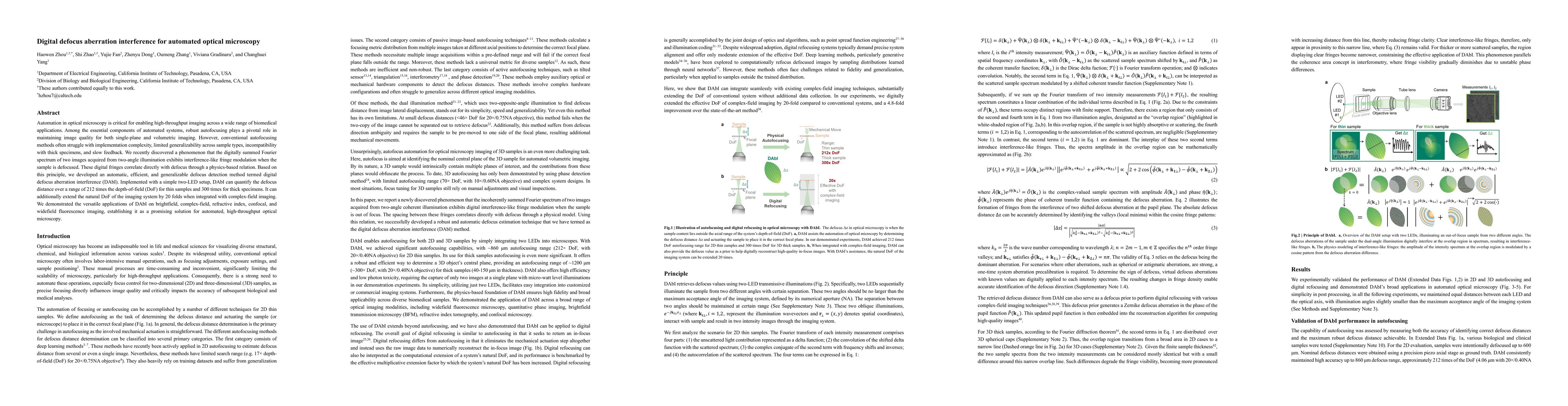
Automation in optical microscopy is critical for enabling high-throughput imaging across a wide range of biomedical applications. Among the essential components of automated systems, robust autofocusing plays a pivotal role in maintaining image quality for both single-plane and volumetric imaging. However, conventional autofocusing methods often struggle with implementation complexity, limited generalizability across sample types, incompatibility with thick specimens, and slow feedback. We recently discovered a phenomenon that the digitally summed Fourier spectrum of two images acquired from two-angle illumination exhibits interference-like fringe modulation when the sample is defocused. These digital fringes correlate directly with defocus through a physics-based relation. Based on this principle, we developed an automatic, efficient, and generalizable defocus detection method termed digital defocus aberration interference (DAbI). Implemented with a simple two-LED setup, DAbI can quantify the defocus distance over a range of 212 times the depth-of-field (DoF) for thin samples and 300 times for thick specimens. It can additionally extend the natural DoF of the imaging system by 20 folds when integrated with complex-field imaging. We demonstrated the versatile applications of DAbI on brightfield, complex-field, refractive index, confocal, and widefield fluorescence imaging, establishing it as a promising solution for automated, high-throughput optical microscopy.
AI Key Findings
Get AI-generated insights about this paper's methodology, results, significance, and more — seven facets brought into focus.
Impact
Authors
PDF Preview
Citation Network
Current paper (gray), citations (green), references (blue)
Display is limited for performance on very large graphs.
Discussion 0