Dilated Convolutional Neural Networks for Cardiovascular MR Segmentation in Congenital Heart Disease
Publication
Metrics
AI Quick Summary
This paper proposes an automatic method using dilated convolutional neural networks (CNNs) for segmenting the myocardium and blood pool in cardiovascular MR (CMR) scans of congenital heart disease patients. The method achieved high accuracy with Dice indices of 0.80±0.06 and 0.93±0.02 for myocardium and blood pool, respectively.
Paper Preview
Abstract
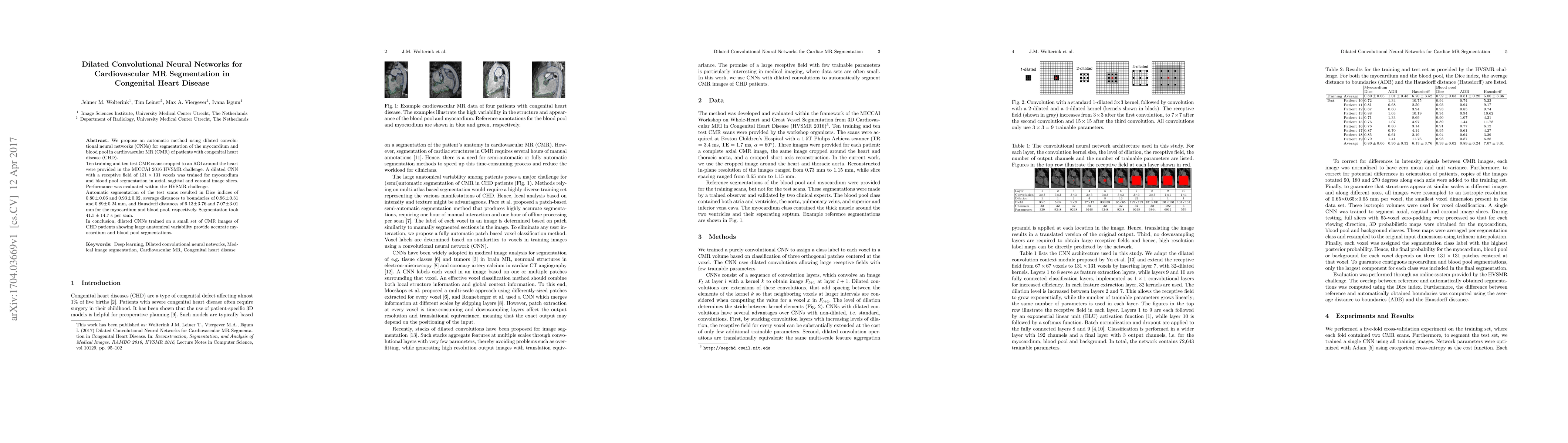
We propose an automatic method using dilated convolutional neural networks (CNNs) for segmentation of the myocardium and blood pool in cardiovascular MR (CMR) of patients with congenital heart disease (CHD). Ten training and ten test CMR scans cropped to an ROI around the heart were provided in the MICCAI 2016 HVSMR challenge. A dilated CNN with a receptive field of 131x131 voxels was trained for myocardium and blood pool segmentation in axial, sagittal and coronal image slices. Performance was evaluated within the HVSMR challenge. Automatic segmentation of the test scans resulted in Dice indices of 0.80$\pm$0.06 and 0.93$\pm$0.02, average distances to boundaries of 0.96$\pm$0.31 and 0.89$\pm$0.24 mm, and Hausdorff distances of 6.13$\pm$3.76 and 7.07$\pm$3.01 mm for the myocardium and blood pool, respectively. Segmentation took 41.5$\pm$14.7 s per scan. In conclusion, dilated CNNs trained on a small set of CMR images of CHD patients showing large anatomical variability provide accurate myocardium and blood pool segmentations.
AI Key Findings
Get AI-generated insights about this paper's methodology, results, significance, and more — seven facets brought into focus.
Impact
Paper Details
PDF Preview
Key Terms
Citation Network
Current paper (gray), citations (green), references (blue)
Display is limited for performance on very large graphs.
Discussion 0