Publication
Metrics
AI Quick Summary
This paper presents a method to directly pattern electrostatic potential on hydroxyapatite coatings using a focused electron beam, creating micro-domains of positive and negative surface potential. The technique's effectiveness is validated by Kelvin Probe Force Microscopy, demonstrating the influence of electron beam charge dose and injection speed on domain shape and potential difference.
Paper Preview
Abstract
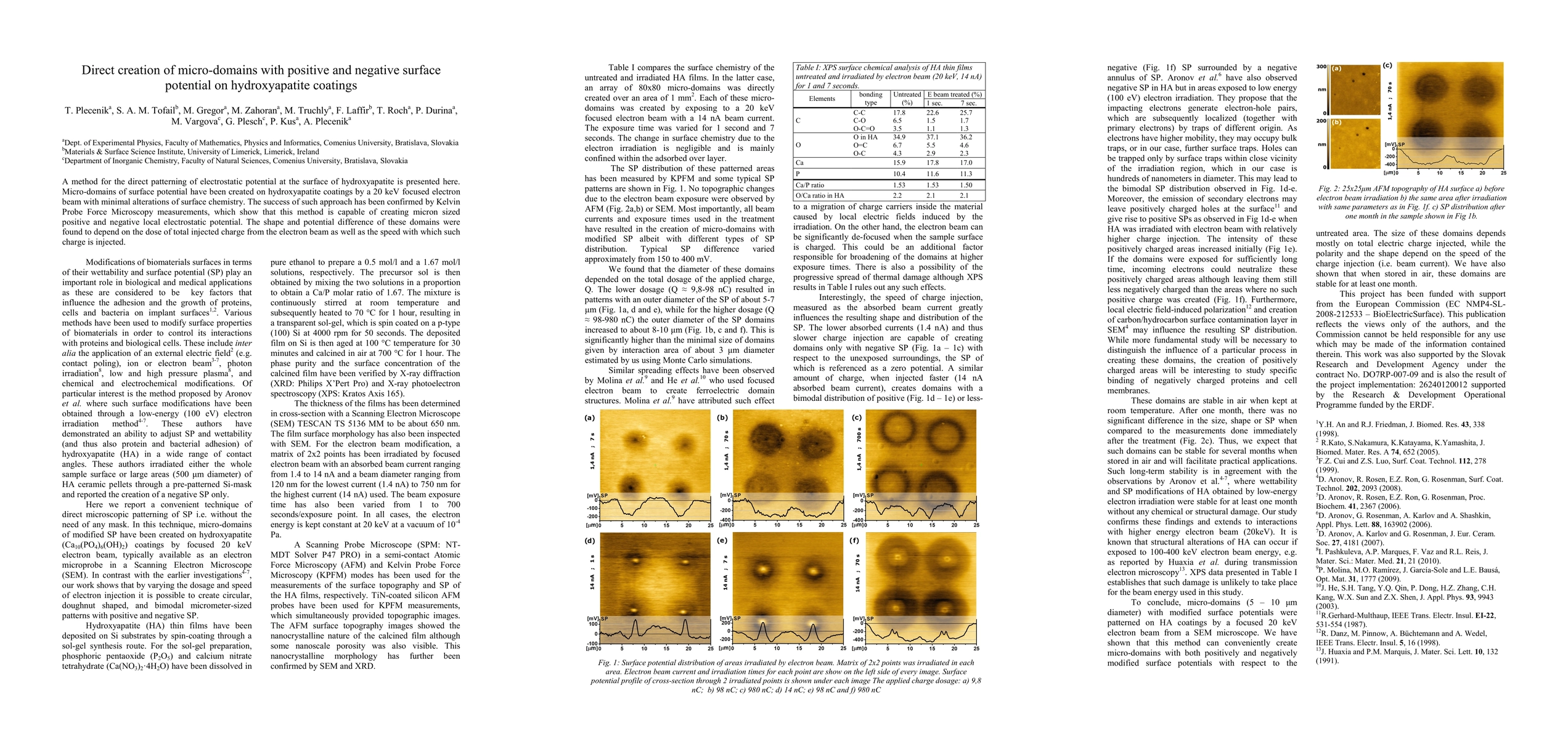
A method for the direct patterning of electrostatic potential at the surface of hydroxyapatite is presented here. Micro-domains of surface potential have been created on hydroxyapatite coatings by a 20 keV focused electron beam with minimal alterations of surface chemistry. The success of such approach has been confirmed by Kelvin Probe Force Microscopy measurements, which show that this method is capable of creating micron sized positive and negative local electrostatic potential. The shape and potential difference of these domains were found to depend on the dose of total injected charge from the electron beam as well as the speed with which such charge is injected.
AI Key Findings
Get AI-generated insights about this paper's methodology, results, significance, and more — seven facets brought into focus.
Impact
Paper Details
PDF Preview
Key Terms
Citation Network
Current paper (gray), citations (green), references (blue)
Display is limited for performance on very large graphs.
Discussion 0