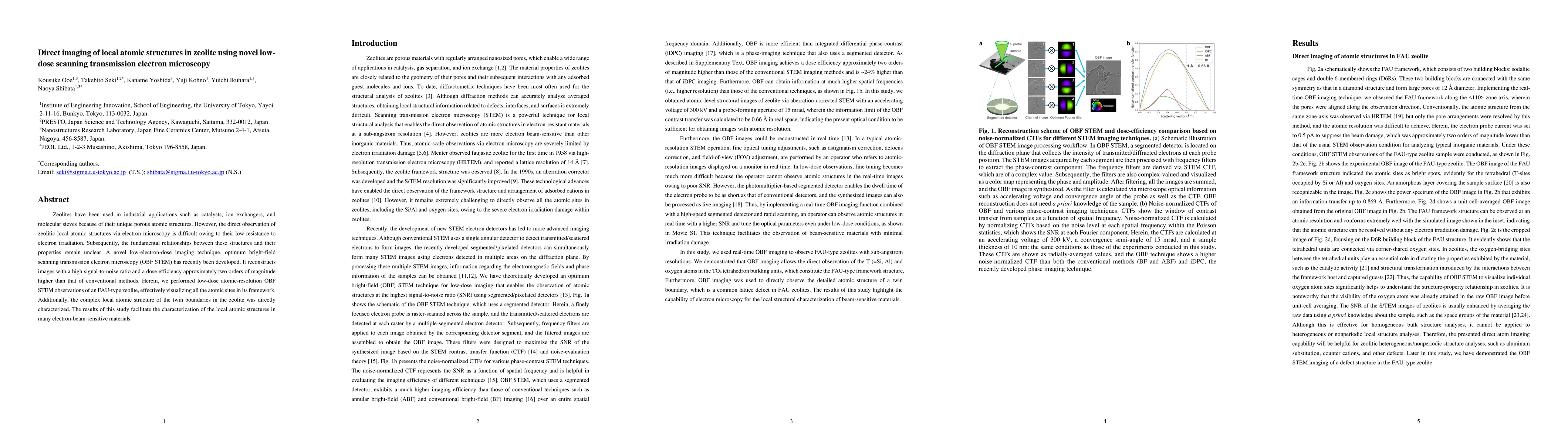
Direct imaging of local atomic structures in zeolite using novel low-dose scanning transmission electron microscopy
Publication
Metrics
AI Quick Summary
The study employs a novel low-dose scanning transmission electron microscopy technique (OBF STEM) to directly image the atomic structures within a FAU-type zeolite, overcoming electron irradiation damage and revealing the complex local atomic structure of its twin boundaries. This advancement allows for better understanding of the relationships between zeolitic atomic structures and their properties.
Paper Preview
Abstract
Zeolites have been used in industrial applications such as catalysts, ion exchangers, and molecular sieves because of their unique porous atomic structures. However, the direct observation of zeolitic local atomic structures via electron microscopy is difficult owing to their low resistance to electron irradiation. Subsequently, the fundamental relationships between these structures and their properties remain unclear. A novel low-electron-dose imaging technique, optimum bright-field scanning transmission electron microscopy (OBF STEM) has recently been developed. It reconstructs images with a high signal-to-noise ratio and a dose efficiency approximately two orders of magnitude higher than that of conventional methods. Herein, we performed low-dose atomic-resolution OBF STEM observations of an FAU-type zeolite, effectively visualizing all the atomic sites in its framework. Additionally, the complex local atomic structure of the twin boundaries in the zeolite was directly characterized. The results of this study facilitate the characterization of the local atomic structures in many electron-beam-sensitive materials.
AI Key Findings
Get AI-generated insights about this paper's methodology, results, significance, and more — seven facets brought into focus.
Impact
Paper Details
Authors
PDF Preview
Key Terms
Citation Network
Current paper (gray), citations (green), references (blue)
Display is limited for performance on very large graphs.
Discussion 0