01
MethodologyHow they did it
Single-molecule imaging technique was used to measure the diffusion of enzymes
This paper uses a novel direct single-molecule imaging technique to observe enhanced enzyme diffusion in the presence of substrates, finding a higher relative increase than previous FCS-based studies. The method reveals that the diffusion enhancement is independent of total enzyme concentration and does not alter enzyme oligomerization state.
This paper uses a novel direct single-molecule imaging technique to observe enhanced enzyme diffusion in the presence of substrates, finding a higher relative increase than previous FCS-based studies. The method reveals that the diffusion enhancement is independent of total enzyme concentration and does not alter enzyme oligomerization state.
Single-molecule imaging technique was used to measure the diffusion of enzymes More in Methodology →
Enhanced diffusion rates observed for urease in the presence of urea — No significant changes in oligomerization state detected More in Key Results →
Understanding the mechanism behind enhanced enzyme diffusion is crucial for biotechnological applications More in Significance →
Limited spatial resolution of single-molecule imaging technique — Potential artifacts from sample preparation or instrumentation More in Limitations →
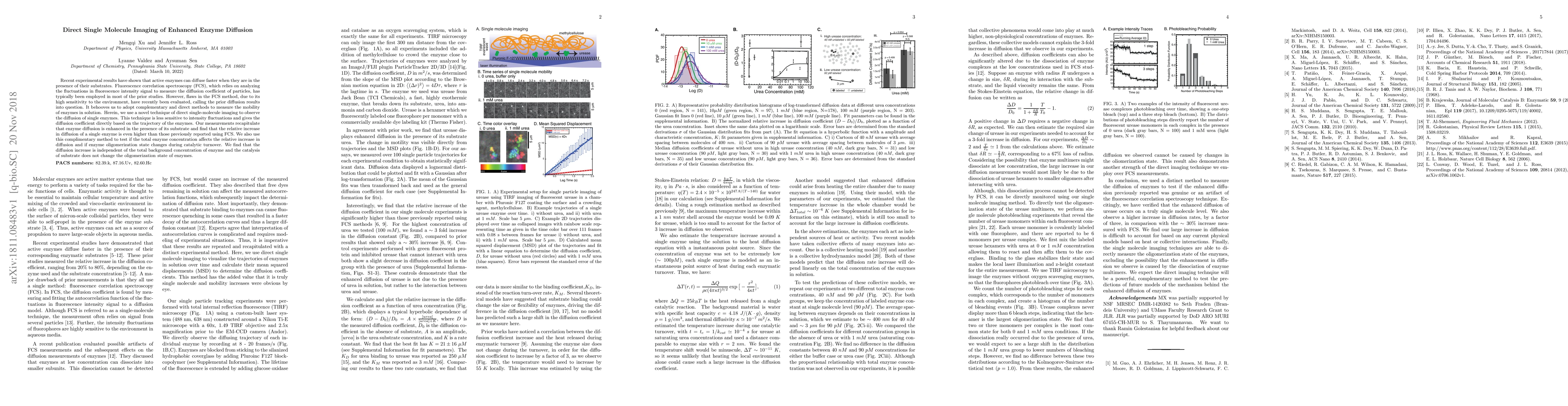
Recent experimental results have shown that active enzymes can diffuse faster when they are in the presence of their substrates. Fluorescence correlation spectroscopy (FCS), which relies on analyzing the fluctuations in fluorescence intensity signal to measure the diffusion coefficient of particles, has typically been employed in most of the prior studies. However, flaws in the FCS method, due to its high sensitivity to the environment, have recently been evaluated, calling the prior diffusion results into question. It behooves us to adopt complimentary and direct methods to measure the mobility of enzymes in solution. Herein, we use a novel technique of direct single-molecule imaging to observe the diffusion of single enzymes. This technique is less sensitive to intensity fluctuations and gives the diffusion coefficient directly based on the trajectory of the enzymes. Our measurements recapitulate that enzyme diffusion is enhanced in the presence of its substrate and find that the relative increase in diffusion of a single enzyme is even higher than those previously reported using FCS. We also use this complementary method to test if the total enzyme concentration affects the relative increase in diffusion and if enzyme oligomerization state changes during catalytic turnover. We find that the diffusion increase is independent of the total background concentration of enzyme and the catalysis of substrate does not change the oligomerization state of enzymes.
Seven facets of this paper, analysed and brought into focus by AI.
Understanding the mechanism behind enhanced enzyme diffusion is crucial for biotechnological applications
Single-molecule imaging technique was used to measure the diffusion of enzymes
Understanding the mechanism behind enhanced enzyme diffusion is crucial for biotechnological applications
Direct observation of enzyme diffusion at the single-molecule level using a custom-built single-molecule imaging setup
The use of single-molecule imaging to study enzyme diffusion and its modulation by urea, providing new insights into the mechanisms underlying enhanced enzyme activity
Current paper (gray), citations (green), references (blue)
Display is limited for performance on very large graphs.
Discussion 0