Publication
Metrics
Quick Actions
AI Quick Summary
A new method uses a fully convolutional autoencoder to identify breast cancer in histopathology images with high accuracy, outperforming existing methods, and provides a visualized probability map for verification.
Quick Answers
What is "Discriminative Pattern Mining for Breast Cancer Histopathology Image Classification via Fully Convolutional Autoencoder" about?
A new method uses a fully convolutional autoencoder to identify breast cancer in histopathology images with high accuracy, outperforming existing methods, and provides a visualized probability map for verification.
What methodology did the authors use?
The paper proposes a discriminative pattern mining approach for invasive carcinoma diagnosis in routine H&E stained breast tissue histopathology images using a fully convolutional autoencoder and one-class SVM. More in Methodology →
What are the key results?
The proposed method outperforms existing patch-based deep learning methods for breast cancer histopathology image classification. — The method identifies suspected regions of malignant cell clusters in images by learning contrast patterns between normal and malignant breast cancer images. More in Key Results →
Why is this work significant?
This research is significant as it presents a practical, generalizable, and self-interpretable method for breast cancer diagnosis, which could facilitate better understanding of the pattern mining and diagnosis solution, and assist pathologists in verifying diagnosis reasoning. More in Significance →
What are the main limitations?
The study's major limitation is the size of the experimental image set and the absence of an external validation group. — The reliance on a single public image set may limit the generalizability of the findings to diverse datasets. More in Limitations →
Abstract
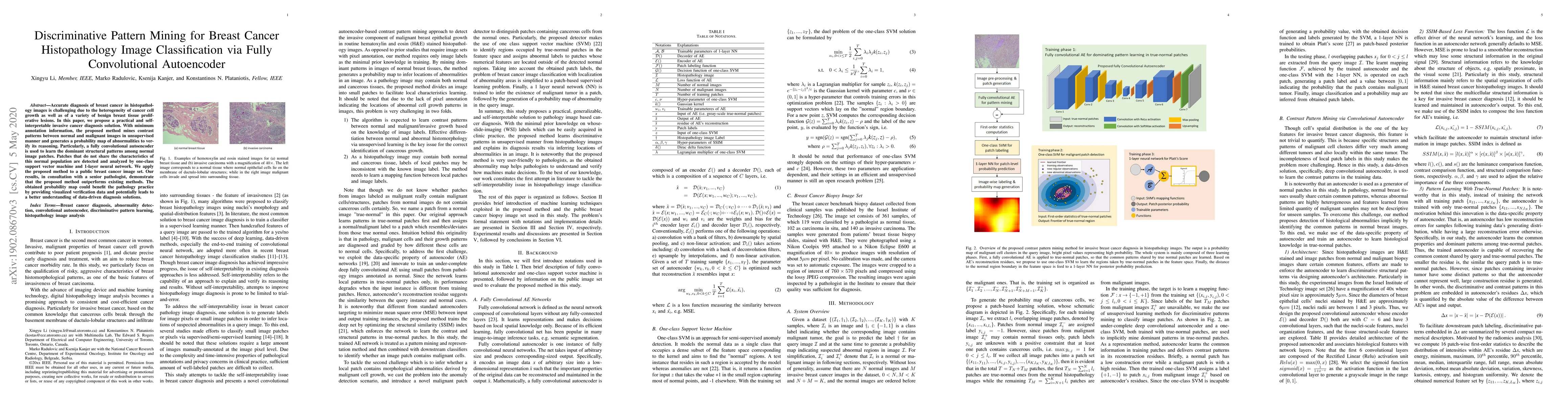
Accurate diagnosis of breast cancer in histopathology images is challenging due to the heterogeneity of cancer cell growth as well as of a variety of benign breast tissue proliferative lesions. In this paper, we propose a practical and self-interpretable invasive cancer diagnosis solution. With minimum annotation information, the proposed method mines contrast patterns between normal and malignant images in unsupervised manner and generates a probability map of abnormalities to verify its reasoning. Particularly, a fully convolutional autoencoder is used to learn the dominant structural patterns among normal image patches. Patches that do not share the characteristics of this normal population are detected and analyzed by one-class support vector machine and 1-layer neural network. We apply the proposed method to a public breast cancer image set. Our results, in consultation with a senior pathologist, demonstrate that the proposed method outperforms existing methods. The obtained probability map could benefit the pathology practice by providing visualized verification data and potentially leads to a better understanding of data-driven diagnosis solutions.
AI Key Findings
Generated Sep 01, 2025
Methodology — What approach did the authors take?
The paper proposes a discriminative pattern mining approach for invasive carcinoma diagnosis in routine H&E stained breast tissue histopathology images using a fully convolutional autoencoder and one-class SVM.
Key Results — What are the main findings?
- The proposed method outperforms existing patch-based deep learning methods for breast cancer histopathology image classification.
- The method identifies suspected regions of malignant cell clusters in images by learning contrast patterns between normal and malignant breast cancer images.
- The obtained probability map provides insights and verification for the image diagnosis result by inferring locations of abnormalities in an image.
- The method requires minimal prior knowledge (WSI labels) and is generalizable to unseen images.
- The approach is self-interpretable, generating visualized information to support and verify its decision.
Significance — Why does this research matter?
This research is significant as it presents a practical, generalizable, and self-interpretable method for breast cancer diagnosis, which could facilitate better understanding of the pattern mining and diagnosis solution, and assist pathologists in verifying diagnosis reasoning.
Technical Contribution — What is the technical contribution?
The main technical contribution is the development of a contrast pattern mining approach using a fully convolutional autoencoder and one-class SVM for breast cancer diagnosis, which is practical, generalizable, and self-interpretable.
Novelty — What is new about this work?
The novelty of this work lies in its practicality, generalizability, and self-interpretability, contrasting with previous deep learning methods that require extensive annotated data and lack transparency in decision-making.
Limitations — What are the limitations of this study?
- The study's major limitation is the size of the experimental image set and the absence of an external validation group.
- The reliance on a single public image set may limit the generalizability of the findings to diverse datasets.
- The method's performance may vary with different staining protocols and image acquisition devices.
Future Work — What did the authors propose for future work?
- Investigate more efficient pattern mining methods to improve diagnosis performance.
- Explore ways to address the black-box property of deep learning in digital pathology.
- Expand the study using larger and more diverse datasets for validation.
Paper Details
How to Cite This Paper
@article{anon2020discriminative,
title = {Discriminative Pattern Mining for Breast Cancer Histopathology Image
Classification via Fully Convolutional Autoencoder},
year = {2020},
eprint = {1902.08670},
archivePrefix = {arXiv},
primaryClass = {cs.CV},
doi = {10.1109/ACCESS.2019.2904245},
}
(2020). Discriminative Pattern Mining for Breast Cancer Histopathology Image
Classification via Fully Convolutional Autoencoder. arXiv. https://doi.org/10.1109/ACCESS.2019.2904245
"Discriminative Pattern Mining for Breast Cancer Histopathology Image
Classification via Fully Convolutional Autoencoder." arXiv, 2020, doi.org/10.1109/ACCESS.2019.2904245.
PDF Preview
Key Terms
Citation Network
Current paper (gray), citations (green), references (blue)
Display is limited for performance on very large graphs.
Similar Papers
Found 4 papersStain Normalized Breast Histopathology Image Recognition using Convolutional Neural Networks for Cancer Detection
Sruthi Krishna, Suganthi S. S, Shivsubramani Krishnamoorthy et al.
Breast Cancer Histopathology Image Classification and Localization using Multiple Instance Learning
Dipesh Tamboli, Abhijeet Patil, Deepak Anand et al.
SHISRCNet: Super-resolution And Classification Network For Low-resolution Breast Cancer Histopathology Image
Xin Zhang, Qingni Shen, Luyuan Xie et al.
| Title | Authors | Year | Actions |
|---|
Comments (0)