Disease Detection in Weakly Annotated Volumetric Medical Images using a Convolutional LSTM Network
Publication
Metrics
AI Quick Summary
This research proposes a convolutional LSTM network for detecting emphysema in low-dose CT lung scans using weakly annotated volumetric medical images. The method effectively learns from large datasets with minimal annotations, achieving an AUC of 0.83 in distinguishing emphysema, outperforming both 2D CNNs and 3D CNNs.
Paper Preview
Abstract
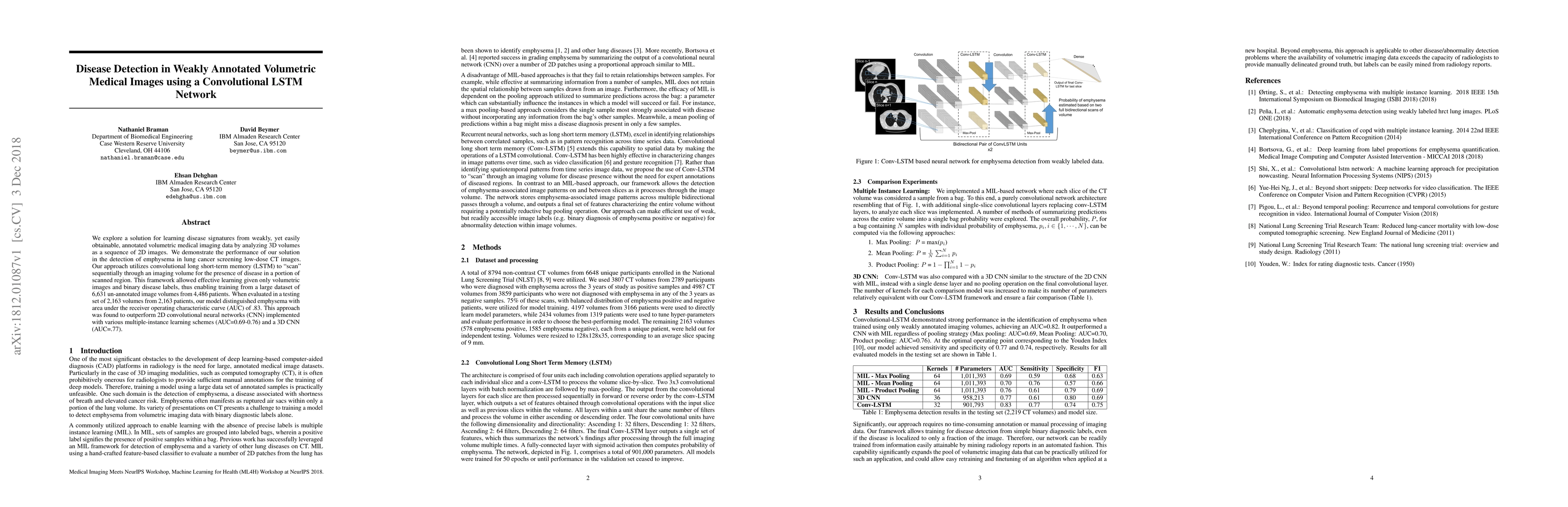
We explore a solution for learning disease signatures from weakly, yet easily obtainable, annotated volumetric medical imaging data by analyzing 3D volumes as a sequence of 2D images. We demonstrate the performance of our solution in the detection of emphysema in lung cancer screening low-dose CT images. Our approach utilizes convolutional long short-term memory (LSTM) to "scan" sequentially through an imaging volume for the presence of disease in a portion of scanned region. This framework allowed effective learning given only volumetric images and binary disease labels, thus enabling training from a large dataset of 6,631 un-annotated image volumes from 4,486 patients. When evaluated in a testing set of 2,163 volumes from 2,163 patients, our model distinguished emphysema with area under the receiver operating characteristic curve (AUC) of .83. This approach was found to outperform 2D convolutional neural networks (CNN) implemented with various multiple-instance learning schemes (AUC=0.69-0.76) and a 3D CNN (AUC=.77).
AI Key Findings
Get AI-generated insights about this paper's methodology, results, significance, and more — seven facets brought into focus.
Impact
Paper Details
Authors
PDF Preview
Key Terms
Citation Network
Current paper (gray), citations (green), references (blue)
Display is limited for performance on very large graphs.
Discussion 0