Summary
Identifying new disease-related patterns in medical imaging data with the help of machine learning enlarges the vocabulary of recognizable findings. This supports diagnostic and prognostic assessment. However, image appearance varies not only due to biological differences, but also due to imaging technology linked to vendors, scanning- or re- construction parameters. The resulting domain shifts impedes data representation learning strategies and the discovery of biologically meaningful cluster appearances. To address these challenges, we introduce an approach to actively learn the domain shift via post-hoc rotation of the data latent space, enabling disentanglement of biological and technical factors. Results on real-world heterogeneous clinical data showcase that the learned disentangled representation leads to stable clusters representing tissue-types across different acquisition settings. Cluster consistency is improved by +19.01% (ARI), +16.85% (NMI), and +12.39% (Dice) compared to the entangled representation, outperforming four state-of-the-art harmonization methods. When using the clusters to quantify tissue composition on idiopathic pulmonary fibrosis patients, the learned profiles enhance Cox survival prediction. This indicates that the proposed label-free framework facilitates biomarker discovery in multi-center routine imaging data. Code is available on GitHub https://github.com/cirmuw/latent-space-rotation-disentanglement.
AI Key Findings
Generated Oct 02, 2025
Methodology
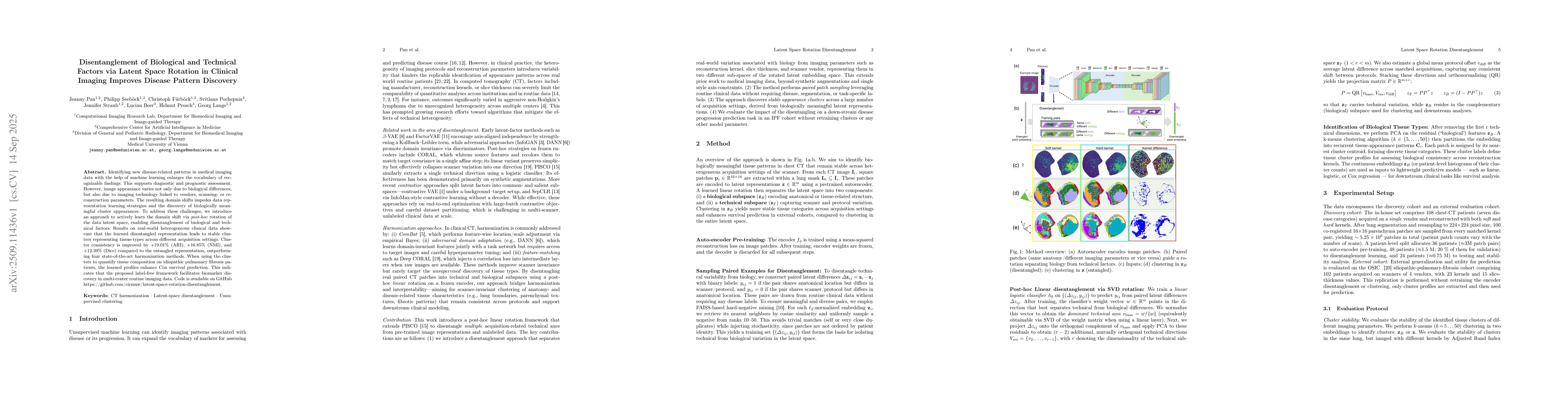
The paper proposes a post-hoc approach to disentangle biological and technical factors in clinical imaging data by rotating the latent space of an autoencoder. This involves training a linear classifier to predict technical differences from paired latent representations, then using singular value decomposition (SVD) to identify and remove technical variation, leaving a biological subspace for clustering and analysis.
Key Results
- Disentangled representations achieved +19.01% improvement in Adjusted Rand Index (ARI), +16.85% in Normalized Mutual Information (NMI), and +12.39% in Dice score compared to entangled representations.
- The method outperformed four state-of-the-art harmonization techniques including DANN, DeepCORAL, β-VAE, and ComBat.
- Disentangled tissue profiles improved Cox survival prediction in idiopathic pulmonary fibrosis (IPF) patients, demonstrating enhanced clinical utility.
Significance
This research enables more reliable biomarker discovery in multi-center clinical imaging data by separating biological signals from technical artifacts. The approach improves the stability and biological relevance of tissue appearance clusters, which is critical for accurate diagnosis and prognosis in heterogeneous medical imaging datasets.
Technical Contribution
The paper introduces a linear post-hoc disentanglement technique using SVD rotation of the latent space, enabling domain shift separation without requiring retraining of the autoencoder or disease-specific supervision.
Novelty
This work presents a novel label-free framework for disentangling biological and technical factors in clinical imaging through latent space rotation, offering a practical solution for harmonizing multi-center, heterogeneous imaging data.
Limitations
- The method assumes paired reconstructions from the same patient, limiting its applicability to unpaired or multi-scanner data.
- The internal cohort includes only two scanners per vendor, which may not capture rare protocol combinations.
Future Work
- Extending the method to handle unpaired or multi-scanner data without requiring explicit pairing.
- Integrating the approach with end-to-end contrastive learning frameworks for more robust representation learning.
- Applying the technique to other medical imaging modalities beyond CT scans.
Paper Details
PDF Preview
Similar Papers
Found 4 papersDisentanglement via Latent Quantization
Chelsea Finn, Jiajun Wu, Kyle Hsu et al.
Evaluating Disentanglement in Generative Models Without Knowledge of Latent Factors
Alexander Cloninger, Gal Mishne, Chester Holtz
Comments (0)