This paper proposes Serial Crystallography to address sample damage in X-ray diffraction microscopy by distributing the total dose over many hydrated macromolecules. It evaluates the required X-ray fluence for achieving a given resolution, confirming the inverse fourth power dependence of exposure time on resolution and indicating that multiple protein beams are needed for sub-nanometer resolution on current synchrotrons, but not on fourth-generation designs.
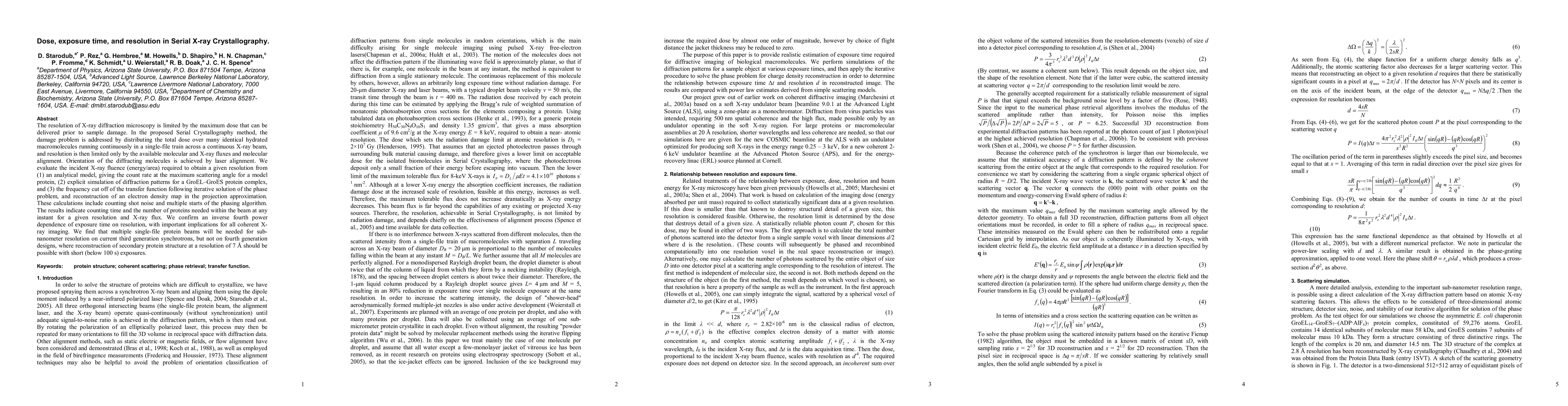
The resolution of X-ray diffraction microscopy is limited by the maximum dose that can be delivered prior to sample damage. In the proposed Serial Crystallography method, the damage problem is addressed by distributing the total dose over many identical hydrated macromolecules running continuously in a single-file train across a continuous X-ray beam, and resolution is then limited only by the available molecular and X-ray fluxes and molecular alignment. Orientation of the diffracting molecules is achieved by laser alignment. We evaluate the incident X-ray fluence (energy/area) required to obtain a given resolution from (1) an analytical model, giving the count rate at the maximum scattering angle for a model protein, (2) explicit simulation of diffraction patterns for a GroEL-GroES protein complex, and (3) the frequency cut off of the transfer function following iterative solution of the phase problem, and reconstruction of an electron density map in the projection approximation. These calculations include counting shot noise and multiple starts of the phasing algorithm. The results indicate counting time and the number of proteins needed within the beam at any instant for a given resolution and X-ray flux. We confirm an inverse fourth power dependence of exposure time on resolution, with important implications for all coherent X-ray imaging. We find that multiple single-file protein beams will be needed for sub-nanometer resolution on current third generation synchrotrons, but not on fourth generation designs, where reconstruction of secondary protein structure at a resolution of 0.7 nm should be possible with short exposures.
Get AI-generated insights about this paper's methodology, results, and significance.
Current paper (gray), citations (green), references (blue)
Wenhui Zhang, Henry N. Chapman, Mansi Butola et al.
Chi Zhang, Ji Liu, Xiaofeng Zhang et al.
Rafal Dunin-Borkowski, Penghan Lu, Sergi Plana-Ruiz et al.
Title | Authors | Year | Actions |
---|---|---|---|
Shielding characteristics of a new epoxy resin reinforced by PbO nanoparticles and PbO micro for protection against X-ray and investigation of their cytotoxicity effects and photocatalytic activity
|
Atefeh Omidtorshiz, M. Benam, M. Momennezhad, Zahra Sabouri, M. Darroudi
|
2024
|
View Paper |
Generation of Large Vortex-Free Superfluid Helium Nanodroplets.
|
A. Ulmer, A. Heilrath, B. Senfftleben, Sean M O O'Connell-Lopez, B. Kruse, L. Seiffert, Katharina Kolatzki, B. Langbehn, A. Hoffmann, T. Baumann, R. Boll, A. Chatterley, A. De Fanis, B. Erk, S. Erukala, A. Feinberg, T. Fennel, P. Grychtol, R. Hartmann, M. Ilchen, M. Izquierdo, B. Krebs, M. Kuster, T. Mazza, Jacobo Montaño, Georg Noffz, D. Rivas, D. Schlosser, F. Seel, H. Stapelfeldt, L. Strüder, J. Tiggesbäumker, H. Yousef, M. Zabel, P. Ziółkowski, M. Meyer, Y. Ovcharenko, A. Vilesov, T. Möller, D. Rupp, R. M. Tanyag
|
2023
|
View Paper |
High-fluence and high-gain multilayer focusing optics to enhance spatial resolution in femtosecond X-ray laser imaging
|
H. Yumoto, T. Koyama, A. Suzuki, Y. Joti, Yoshiya Niida, K. Tono, Yoshitaka Bessho, M. Yabashi, Y. Nishino, H. Ohashi
|
2022
|
View Paper |
Coherent and incoherent imaging of biological specimens with electrons and X-rays.
|
P. Rez
|
2021
|
View Paper |
Calculating absorption dose when X-ray irradiation modifies material quantity and chemistry.
|
V. Berejnov, B. Rubinstein, Lis G. A. Melo, A. Hitchcock
|
2021
|
View Paper |
Relative merits and limiting factors for x-ray and electron microscopy of thick, hydrated organic materials.
|
Ming Du, C. Jacobsen
|
2020
|
View Paper |
The achievable resolution for X-ray imaging of cells and other soft biological material
|
C. Nave
|
2020
|
View Paper |
Admissible Measurements and Robust Algorithms for Ptychography
|
Brian Preskitt, Rayan Saab
|
2019
|
View Paper |
A comparison of absorption and phase contrast for X-ray imaging of biological cells
|
C. Nave
|
2018
|
View Paper |
Anisotropic nano-scale resolution in 3D Bragg coherent diffraction imaging
|
M. Cherukara, W. Cha, R. Harder
|
2018
|
View Paper |
In situ coherent X-ray diffraction imaging of radiation-induced mass loss in metal-polymer composite spheres.
|
E. T. B. Skjønsfjell, Y. Chushkin, F. Zontone, D. Breiby
|
2018
|
View Paper |
Current Status of Single Particle Imaging with X-ray Lasers
|
Zhibin Sun, Jiadong Fan, Haoyuan Li, Huaidong Jiang
|
2018
|
View Paper |
Advanced Photon Source Upgrade Project preliminary design report
|
T. Fornek
|
2017
|
View Paper |
XFELs for structure and dynamics in biology
|
J. Spence
|
2017
|
View Paper |
Enhancing resolution in coherent x-ray diffraction imaging
|
D. Noh, Chan Kim, Yoonhee Kim, Changyong Song
|
2016
|
View Paper |
Non-destructive phase contrast hard x-ray imaging to reveal the three-dimensional microstructure of soft and hard tissues
|
A. Khimchenko, G. Schulz, H. Deyhle, S. Hieber, Samiul Hasan, C. Bikis, J. Schulz, L. Costeur, B. Müller
|
2016
|
View Paper |
Coherent diffractive imaging: towards achieving atomic resolution.
|
S. Dietze, O. Shpyrko
|
2015
|
View Paper |
Signal enhancement and Patterson-search phasing for high-spatial-resolution coherent X-ray diffraction imaging of biological objects
|
Y. Takayama, S. Maki-Yonekura, Tomotaka Oroguchi, M. Nakasako, K. Yonekura
|
2015
|
View Paper |
Real-time and Sub-wavelength Ultrafast Coherent Diffraction Imaging in the Extreme Ultraviolet
|
M. Zürch, J. Rothhardt, S. Hädrich, S. Demmler, M. Krebs, J. Limpert, A. Tünnermann, A. Guggenmos, U. Kleineberg, C. Spielmann
|
2014
|
View Paper |
Cyanobacterial Photosystem II in a native-like superstructure at 2.44 Å resolution and first serial femtosecond X-ray studies
|
J. Hellmich
|
2014
|
View Paper |
Variability of in vivo fluid dose distribution in mouse airways is visualized by high-speed synchrotron X-ray imaging.
|
M. Donnelley, K. Morgan, K. Siu, D. Parsons
|
2013
|
View Paper |
Upsampling speckle patterns for coherent X-ray diffraction imaging
|
Y. Chushkin, F. Zontone
|
2013
|
View Paper |
X-ray free electron lasers motivate bioanalytical characterization of protein nanocrystals: serial femtosecond crystallography.
|
M. Bogan
|
2013
|
View Paper |
Structure determination through correlated fluctuations in x-ray scattering
|
R. Kirian
|
2012
|
View Paper |
X-ray lasers for structural and dynamic biology
|
J. Spence, J. Spence, U. Weierstall, U. Weierstall, H. Chapman
|
2012
|
View Paper |
Room temperature femtosecond X-ray diffraction of photosystem II microcrystals
|
J. Kern, R. Alonso-Mori, J. Hellmich, Rosalie Tran, J. Hattne, H. Laksmono, Carina Glöckner, N. Echols, R. Sierra, J. Sellberg, B. Lassalle‐Kaiser, R. Gildea, P. Glatzel, R. Grosse-Kunstleve, M. Latimer, Trevor A. McQueen, D. Difiore, A. Fry, M. Messerschmidt, A. Miahnahri, D. Schafer, M. Seibert, D. Sokaras, T. Weng, P. Zwart, W. White, P. Adams, M. Bogan, S. Boutet, Garth J. Williams, J. Messinger, N. Sauter, A. Zouni, U. Bergmann, J. Yano, V. Yachandra
|
2012
|
View Paper |
Energy recovery linac (ERL) coherent hard x-ray sources
|
D. Bilderback, J. Brock, D. Dale, K. Finkelstein, M. Pfeifer, S. Gruner
|
2010
|
View Paper |
The minimum crystal size needed for a complete diffraction data set
|
J. Holton, K. Frankel
|
2010
|
View Paper |
Dose requirements for resolving a given feature in an object by coherent x-ray diffraction imaging
|
A. Schropp, C. Schroer
|
2010
|
View Paper |
Hard X-ray Microscopy with Elemental, Chemical and Structural Contrast
|
C. Schroer, P. Boye, J. Feldkamp, J. Patommel, A. Schropp, D. Samberg, S. Stephan, M. Burghammer, S. Schöder, C. Riekel, B. Lengeler, G. Falkenberg, G. Wellenreuther, M. Kuhlmann, R. Frahm, D. Lützenkirchen-Hecht, W. Schroeder
|
2010
|
View Paper |
SEM imaging of liquid jets.
|
D. DePonte, R. Doak, M. Hunter, Zhenquan Liu, U. Weierstall, J. Spence
|
2009
|
View Paper |
Feasibility of imaging living cells at subnanometer resolutions by ultrafast X-ray diffraction
|
M. Bergh, G. Huldt, N. Tîmneanu, F. Maia, J. Hajdu
|
2008
|
View Paper |
Powder diffraction from a continuous microjet of submicrometer protein crystals.
|
D. Shapiro, D. Shapiro, H. Chapman, D. DePonte, R. Doak, P. Fromme, G. Hembree, Mark S. Hunter, S. Marchesini, S. Marchesini, K. Schmidt, J. Spence, D. Starodub, U. Weierstall
|
2008
|
View Paper |
Ultrafast soft X-ray scattering and reference-enhanced diffractive imaging of weakly scattering nanoparticles
|
S. Boutet, M. Bogan, A. Barty, M. Frank, W. H. Benner, S. Marchesini, M. Seibert, J. Hajdu, Henry N. Chapman
|
2008
|
View Paper |
Physical optics basis for the X-ray phase imaging of low contrast samples: a primer
|
A. Peele, K. Nugent
|
2008
|
View Paper |
Experimental approaches for solution X-ray scattering and fiber diffraction.
|
H. Tsuruta, T. Irving
|
2008
|
View Paper |
Gas dynamic virtual nozzle for generation of microscopic droplet streams
|
D. DePonte, U. Weierstall, K. Schmidt, J. Warner, D. Starodub, J. Spence, R. Doak
|
2008
|
View Paper |
An assessment of the resolution limitation due to radiation-damage in x-ray diffraction microscopy.
|
M. Howells, T. Beetz, H. Chapman, C. Cui, J. Holton, J. Holton, C. Jacobsen, C. Jacobsen, J. Kirz, J. Kirz, E. Lima, S. Marchesini, H. Miao, D. Sayre, D. Shapiro, J. Spence, J. Spence, D. Starodub
|
2005
|
View Paper |
Direct structure-determination of nodavirus in solution by x-ray ensemble diffraction microscopy
|
Ning-Jung Chen, Chia-Hui Yeh, Huai-Yu Cao, Nai-Chi Chen, Chun-Jung Chen, Yi-Wei Tsai, C. Chen, Yu-Shan Huang, Chien-Chun Chen, Jhih-Heng Yang, Jhih-Min Lin
|
2023
|
View Paper |
A Historical Review of the Structures of Water and Ice
|
V. Artemov
|
2021
|
View Paper |
Diffractive Imaging of Single Particles
|
J. Spence
|
2019
|
View Paper |
Processing of Diffraction Patterns Obtained from X-Ray Diffraction Imaging Experiments Using X-Ray Free Electron Laser Pulses
|
M. Nakasako
|
2018
|
View Paper |
X-Ray Lasers in Biology: Structure and Dynamics
|
J. Spence
|
2017
|
View Paper |
Dose requirements for resolving a given feature in an object by coherent x-ray diffraction imaging
|
A. Diaz, Garth J. Williams
|
2010
|
View Paper |
New Journal of Physics Energy recovery linac ( ERL ) coherent hard x-ray sources
|
D. Bilderback, J. Brock, D. Dale, K. Finkelstein, M. Pfeifer, S. Gruner
|
2010
|
View Paper |
SCIENCE CASE FOR A NEW LIGHT SOURCE (NLS)
|
A. Cavalleri, J. Underwood, F. Quinn
|
2008
|
View Paper |
Feasibilityof imaging living cells at subnanometer resolutionsbyultrafastX-raydiffraction
|
M. Bergh, G. Huldt, N. Tîmneanu, F. Maia, J. Hajdu
|
2008
|
View Paper |
Comments (0)