Druse-Induced Morphology Evolution in Retinal Pigment Epithelium
Publication
Metrics
AI Quick Summary
Researchers studied the effects of soft drusen on retinal pigment epithelium morphology using a combination of experimental and modeling approaches, finding that a purse-string mechanism can explain RPE healing after cell loss due to drusen damage.
Paper Preview
Abstract
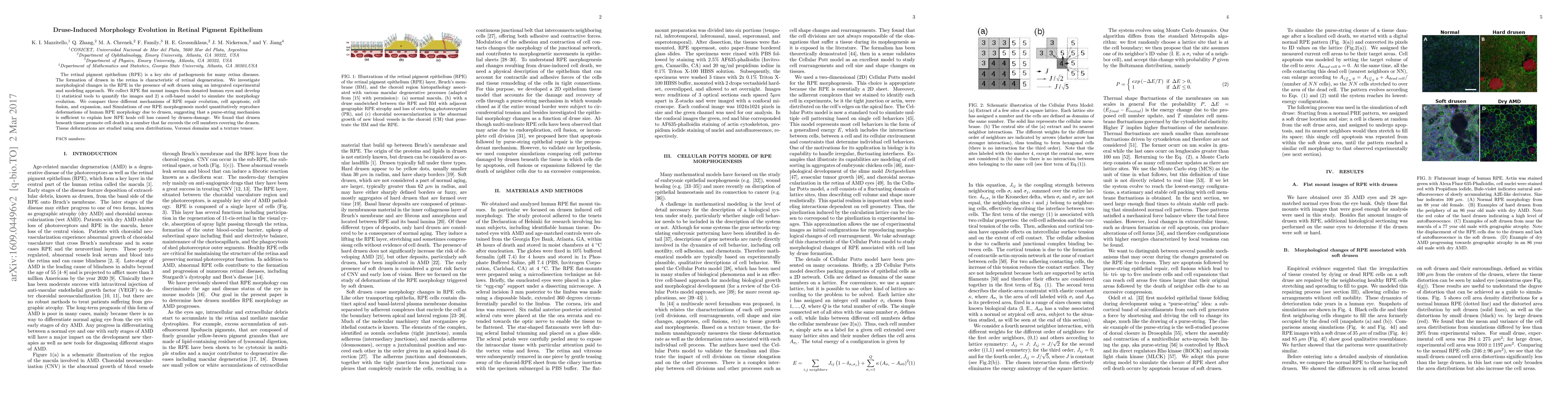
The retinal pigment epithelium (RPE) is a key site of pathogenesis for many retina diseases. The formation of drusen in the retina is characteristic of retinal degeneration. We investigate morphological changes in the RPE in the presence of soft drusen using an integrated experimental and modeling approach. We collect RPE flat mount images from donated human eyes and develop 1) statistical tools to quantify the images and 2) a cell-based model to simulate the morphology evolution. We compare three different mechanisms of RPE repair evolution, cell apoptosis, cell fusion, and expansion, and Simulations of our RPE morphogenesis model quantitatively reproduce deformations of human RPE morphology due to drusen, suggesting that a purse-string mechanism is sufficient to explain how RPE heals cell loss caused by drusen-damage. We found that drusen beneath tissue promote cell death in a number that far exceeds the cell numbers covering the drusen. Tissue deformations are studied using area distributions, Voronoi domains and a texture tensor.
AI Key Findings
Get AI-generated insights about this paper's methodology, results, significance, and more — seven facets brought into focus.
Impact
Paper Details
PDF Preview
Key Terms
Citation Network
Current paper (gray), citations (green), references (blue)
Display is limited for performance on very large graphs.
Discussion 0