ArxivLens

Publication

Published: Aug 04, 2026
Categories: cs.CV, cs.LG

Metrics

Source: ArXiv

Quick Answers

What methodology did the authors use?

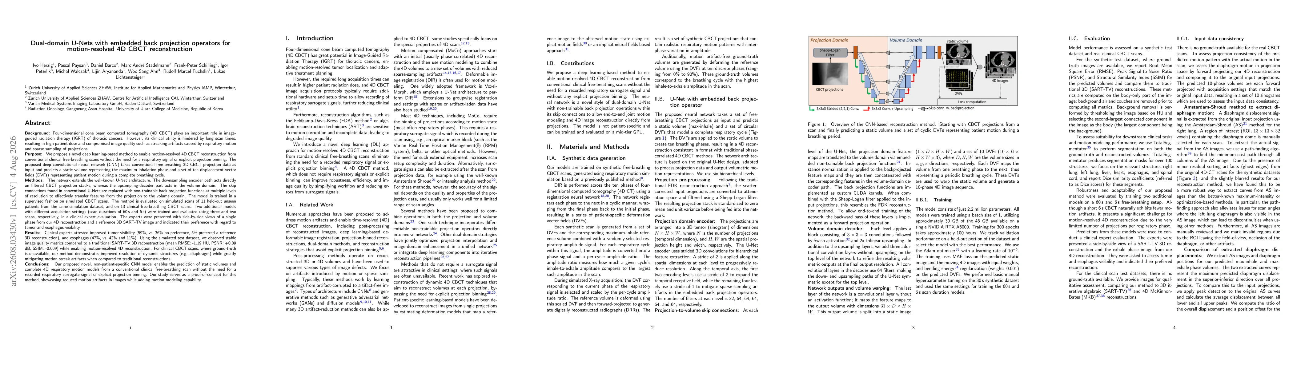
A dual-domain U-Net with non-trainable back-projection operators transfers features between projection and volume domains. The encoder processes filtered CBCT projection stacks, the decoder operates in the volume domain, and skip connections are replaced by back-projection layers. Training is supervised on simulated CBCT data to predict a static maximum-inhalation volume plus ten DVFs representing the breathing cycle, enabling motion-resolved 4D CBCT... More in Methodology →

What are the key results?

Clinical experts preferred the proposed method for tumor visibility in 59% of cases versus 36% for the reference, and for esophagus visibility in 47% versus 42% (with 11% preferring the reference). — On simulated data, image quality matched a traditional 3D SART-TV reconstruction (mean RMSE about -1.19 HU, PSNR +0.09 dB, SSIM -0.009) while enabling 4D reconstruction. More in Key Results →

Why is this work significant?

The approach enables motion-resolved 4D CBCT from a single free-breathing scan without external respiratory signals or projection binning, improving robustness to motion and offering explicit motion-modeling via predicted DVFs. More in Significance →

What are the main limitations?

Evaluation on unseen data relies on simulated training and a limited clinical sample; generalization to broader patient populations remains unverified. — Non-patient-specific training may limit adaptability to atypical motion patterns or severe artifacts not represented in the training set. More in Limitations →

Paper Preview

Abstract

Four-dimensional cone beam CT (4D CBCT) is important for image-guided radiation therapy of thoracic cancers, but its use is limited by long scan times, causing high patient dose and motion/sparse-sampling artifacts. We propose a deep learning method for motion-resolved 4D CBCT reconstruction from conventional free-breathing scans, without a respiratory signal or explicit projection binning. Our CNN takes free-breathing 3D CBCT projections as input and predicts a static volume at maximum inhalation plus ten displacement vector fields (DVFs) spanning a breathing cycle. The network extends U-Net: the encoder acts on filtered projection stacks, the decoder acts in the volume domain, and skip connections are replaced with non-trainable back-projection functions at multiple resolutions to transfer features between domains. The model is trained on simulated CBCT scans and evaluated on 11 unseen simulated patients and 13 clinical free-breathing scans. Two additional models (60 s and 6 s scans) were evaluated by clinical experts on three and two scans, comparing single phases of our 4D reconstruction to reference 3D SART-TV images for tumor and esophagus visibility. Experts preferred our method for tumor visibility (59% vs. 36% no preference, 5% reference) and esophagus visibility (47% vs. 42%, 11%). On simulated data, image quality matched SART-TV (mean RMSE: -1.19 HU, PSNR: +0.09 dB, SSIM: -0.009) while enabling 4D reconstruction. On clinical scans, our method showed sharper dynamic structures (e.g., diaphragm) and fewer motion streak artifacts than traditional reconstruction. This non-patient-specific CNN predicts static volumes and full 4D respiratory motion models from a single free-breathing scan, without a respiratory surrogate or projection binning, reducing motion artifacts while adding motion-modeling capability.
Key Findings, in focus

Seven facets of this paper, analysed and brought into focus by AI.

AI-generated · verify against source Generated Aug 06, 2026
In focus · one-line synthesis

The approach enables motion-resolved 4D CBCT from a single free-breathing scan without external respiratory signals or projection binning, improving robustness to motion and offering explicit motion-modeling via predicted DVFs.

01
MethodologyHow they did it

A dual-domain U-Net with non-trainable back-projection operators transfers features between projection and volume domains. The encoder processes filtered CBCT projection stacks, the decoder operates in the volume domain, and skip connections are replaced by back-projection layers. Training is supervised on simulated CBCT data to predict a static maximum-inhalation volume plus ten DVFs representing the breathing cycle, enabling motion-resolved 4D CBCT without respiratory surrogates or projection binning.

02
ResultsWhat they found
  • Clinical experts preferred the proposed method for tumor visibility in 59% of cases versus 36% for the reference, and for esophagus visibility in 47% versus 42% (with 11% preferring the reference).
  • On simulated data, image quality matched a traditional 3D SART-TV reconstruction (mean RMSE about -1.19 HU, PSNR +0.09 dB, SSIM -0.009) while enabling 4D reconstruction.
  • For clinical CBCT scans, the method produced sharper dynamic structures (e.g., diaphragm) and reduced motion streak artifacts compared to conventional reconstructions.
03
SignificanceWhy it matters

The approach enables motion-resolved 4D CBCT from a single free-breathing scan without external respiratory signals or projection binning, improving robustness to motion and offering explicit motion-modeling via predicted DVFs.

04
Technical ContributionsWhat's new technically

Introduction of a dual-domain CNN architecture that replaces skip connections with non-trainable back-projection functions, enabling end-to-end learning of motion-resolved 4D CBCT from free-breathing projections without respiratory surrogates or projection binning.

05
NoveltyWhat sets it apart

First demonstration of embedding back-projection operators within a U-Net to transfer features across projection and volume domains for 4D CBCT reconstruction, achieving motion modeling from a single free-breathing scan without external surrogate data.

06
LimitationsWhat to be cautious about
  • Evaluation on unseen data relies on simulated training and a limited clinical sample; generalization to broader patient populations remains unverified.
  • Non-patient-specific training may limit adaptability to atypical motion patterns or severe artifacts not represented in the training set.
07
Future WorkWhere it goes next
  • Extend evaluation to larger, multi-institution clinical datasets to validate robustness across scanners and protocols.
  • Investigate integration with real-time workflows and explore improvements in DVF regularization for more accurate motion modeling.
  • Explore ablation studies on back-projection operator placements and multi-resolution effects to further enhance 4D reconstruction quality.

Paper Details

Paper ID: 2608.03430
Comments: 15 pages, 9 Figures
Categories:
cs.CV cs.LG

How to Cite This Paper

Reference manager: RIS EndNote


Discussion 0

This website uses cookies to ensure you get the best experience. Learn more