Dual-Energy Cone-Beam CT Using Two Complementary Limited-Angle Scans with A Projection-Consistent Diffusion Model
Publication
Metrics
AI Quick Summary
This study proposes a method for single-scan dual-energy imaging on cone-beam CT (CBCT) scanners using two complementary limited-angle scans and a projection-consistent diffusion model, achieving quantitative dual-energy projections without hardware modifications. The method showed promising results with mean absolute errors of 20 HU in simulations and 25 HU in real rat data.
Paper Preview
Abstract
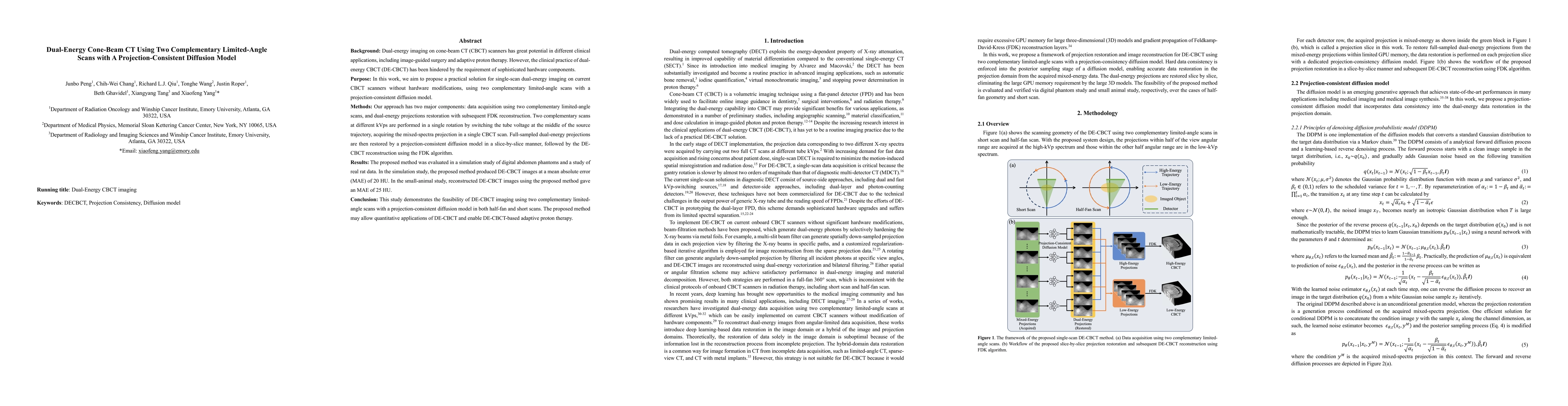
Background: Dual-energy imaging on cone-beam CT (CBCT) scanners has great potential in different clinical applications, including image-guided surgery and adaptive proton therapy. However, the clinical practice of dual-energy CBCT (DE-CBCT) has been hindered by the requirement of sophisticated hardware components. Purpose: In this work, we aim to propose a practical solution for single-scan dual-energy imaging on current CBCT scanners without hardware modifications, using two complementary limited-angle scans with a projection-consistent diffusion model. Methods: Our approach has two major components: data acquisition using two complementary limited-angle scans, and dual-energy projections restoration with subsequent FDK reconstruction. Two complementary scans at different kVps are performed in a single rotation by switching the tube voltage at the middle of the source trajectory, acquiring the mixed-spectra projection in a single CBCT scan. Full-sampled dual-energy projections are then restored by a projection-consistent diffusion model in a slice-by-slice manner, followed by the DE-CBCT reconstruction using the FDK algorithm. Results: The proposed method was evaluated in a simulation study of digital abdomen phantoms and a study of real rat data. In the simulation study, the proposed method produced DE-CBCT images at a mean absolute error (MAE) of 20 HU. In the small-animal study, reconstructed DE-CBCT images using the proposed method gave an MAE of 25 HU. Conclusion: This study demonstrates the feasibility of DE-CBCT imaging using two complementary limited-angle scans with a projection-consistent diffusion model in both half-fan and short scans. The proposed method may allow quantitative applications of DE-CBCT and enable DE-CBCT-based adaptive proton therapy.
AI Key Findings
Get AI-generated insights about this paper's methodology, results, significance, and more — seven facets brought into focus.
Impact
Paper Details
Authors
PDF Preview
Key Terms
Citation Network
Current paper (gray), citations (green), references (blue)
Display is limited for performance on very large graphs.
Discussion 0