The research employs a hybrid CNN-transformer architecture combined with advanced upsampling techniques to enhance medical image segmentation. It utilizes datasets like Kvasir-seg and ISIC for training and evaluation, incorporating deep regression of distance maps and content-aware feature reassembly.
DyGLNet: Hybrid Global-Local Feature Fusion with Dynamic Upsampling for Medical Image Segmentation
Publication
Metrics
Quick Answers
What methodology did the authors use?
The research employs a hybrid CNN-transformer architecture combined with advanced upsampling techniques to enhance medical image segmentation. It utilizes datasets like Kvasir-seg and ISIC for training and evaluation, incorporating deep regression of distance maps and content-aware feature reassembly. More in Methodology →
What are the key results?
Achieved state-of-the-art performance on polyp segmentation tasks with significant improvements in Dice coefficients. — Demonstrated superior accuracy in skin lesion segmentation compared to existing methods, particularly in handling low-quality images. More in Key Results →
Why is this work significant?
This research advances medical imaging analysis by providing more accurate and efficient segmentation tools, which can improve diagnostic accuracy and treatment planning in clinical settings. More in Significance →
What are the main limitations?
Requires substantial computational resources for training transformer-based models. — Performance may degrade with highly variant or noisy medical imaging data. More in Limitations →
Abstract
Medical image segmentation grapples with challenges including multi-scale lesion variability, ill-defined tissue boundaries, and computationally 
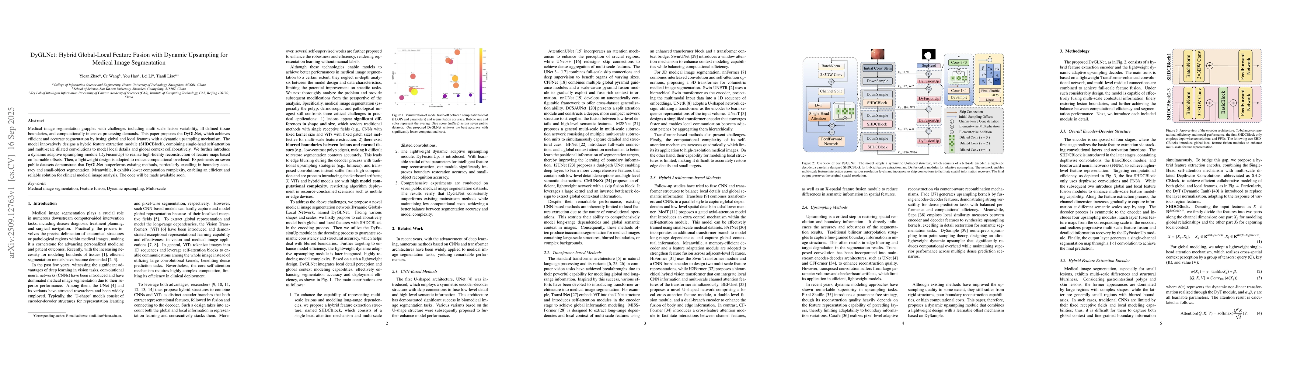
intensive processing demands. This paper proposes the DyGLNet, which achieves efficient and accurate segmentation by fusing global and local features with a dynamic upsampling mechanism. The model innovatively designs a hybrid feature extraction module (SHDCBlock), combining single-head self-attention and multi-scale dilated convolutions to model local details and global context collaboratively. We further introduce a dynamic adaptive upsampling module (DyFusionUp) to realize high-fidelity reconstruction of feature maps based on learnable offsets. Then, a lightweight design is adopted to reduce computational overhead. Experiments on seven public datasets demonstrate that DyGLNet outperforms existing methods, particularly excelling in boundary accuracy and small-object segmentation. Meanwhile, it exhibits lower computation complexity, enabling an efficient and reliable solution for clinical medical image analysis. The code will be made available soon.
Key Findings, in focus
Seven facets of this paper, analysed and brought into focus by AI.
This research advances medical imaging analysis by providing more accurate and efficient segmentation tools, which can improve diagnostic accuracy and treatment planning in clinical settings.
- Achieved state-of-the-art performance on polyp segmentation tasks with significant improvements in Dice coefficients.
- Demonstrated superior accuracy in skin lesion segmentation compared to existing methods, particularly in handling low-quality images.
- Enabled real-time processing capabilities through efficient sub-pixel convolutional networks and transformer-based encoders.
This research advances medical imaging analysis by providing more accurate and efficient segmentation tools, which can improve diagnostic accuracy and treatment planning in clinical settings.
Proposed a hybrid CNN-transformer architecture with innovative upsampling techniques, including similarity-aware point affiliation and content-aware feature reassembly, to enhance segmentation accuracy and efficiency.
This work introduces a novel hybrid architecture that combines the strengths of CNNs and transformers while incorporating advanced upsampling methods, offering significant improvements over traditional segmentation approaches.
- Requires substantial computational resources for training transformer-based models.
- Performance may degrade with highly variant or noisy medical imaging data.
- Exploring lightweight transformer variants for mobile and edge computing applications.
- Integrating multi-modal data (e.g., histopathology and imaging) for more comprehensive analysis.
- Developing adaptive architectures that can handle diverse imaging modalities and pathologies.
Discussion 0