Publication
Metrics
AI Quick Summary
This paper introduces dynamic full-field optical coherence tomography for non-invasive 3D live-imaging of human retinal organoids, providing functional insights through organelle motility with sub-micrometer spatial and millisecond temporal resolution. This technique enables identification of specific cell types based on their functional characteristics.
Paper Preview
Abstract
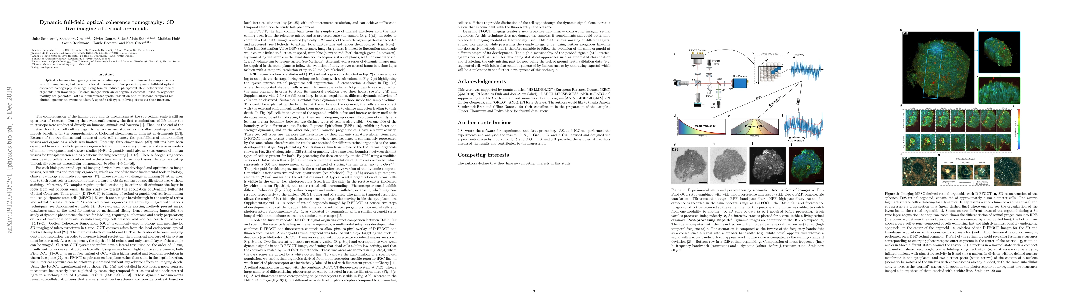
Optical coherence tomography offers astounding opportunities to image the complex structure of living tissue, but lacks functional information. We present dynamic full-field optical coherence tomography to image living human induced pluripotent stem cell-derived retinal organoids non-invasively. Colored images with an endogenous contrast linked to organelle motility are generated, with sub-micrometer spatial resolution and millisecond temporal resolution, opening an avenue to identify specific cell types in living tissue via their function.
AI Key Findings
Get AI-generated insights about this paper's methodology, results, significance, and more — seven facets brought into focus.
Impact
Paper Details
Authors
PDF Preview
Key Terms
Citation Network
Current paper (gray), citations (green), references (blue)
Display is limited for performance on very large graphs.
Discussion 0