Dynamic radiological anthropomorphic thoracic phantom with a deformable chest wall
Publication
Metrics
AI Quick Summary
This study developed a dynamic radiological anthropomorphic thoracic phantom with a deformable chest wall for radiotherapy dose distribution validation. The phantom, created using a 3D printing-like technique and a breathing mechanism, showed appropriate motion amplitudes during quiet breathing but required manual intervention for deep breathing. Further optimizations are needed for better reproducibility and more realistic tumor displacement.
Paper Preview
Abstract
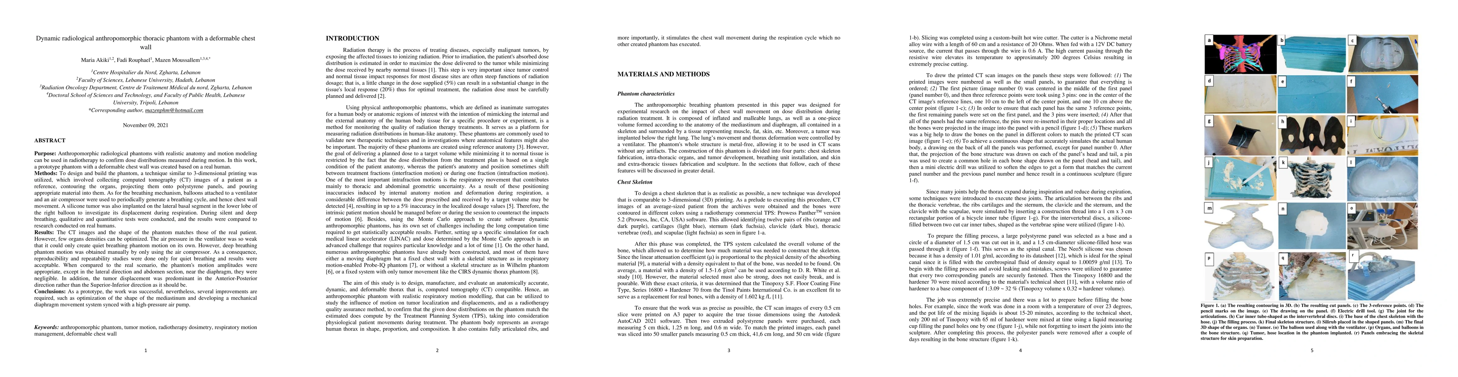
Purpose: Anthropomorphic phantoms can be used in radiotherapy to confirm dose distributions. In this work, a prototype phantom with a deformable chest wall was created based on real human. Methods: A technique similar to 3-dimensional printing was utilized, which involved collecting computed tomography (CT) images of a patient as a reference, contouring the organs, projecting them onto polystyrene panels, and pouring appropriate material into them. Balloons attached to a ventilator and an air compressor were used to periodically generate a breathing cycle. A silicone tumor was also implanted in the lower lobe of the right balloon to investigate its displacement during respiration. During silent and deep breathing, qualitative and quantitative tests were conducted, and the results were compared to research conducted on real humans. Results: The CT images and the shape of the phantom matches those of the real patient. However, few organs densities can be optimized. The air pressure in the ventilator was so weak that it could only create quiet breathing phantom motion on its own. However, deep breathing phantom motion was obtained manually by only using the air compressor. As a consequence, reproducibility and repeatability studies were done only for quiet breathing and results were acceptable. When compared to the real scenario, the phantom's motion amplitudes were appropriate, except in the lateral direction and abdomen section, near the diaphragm, they were negligible. In addition, the tumor displacement was predominant in the Anterior-Posterior direction rather than the Superior-Inferior direction as it should be. Conclusions: As a prototype, the work was successful, nevertheless, several improvements are required, such as optimization of the shape of the mediastinum and developing a mechanical diaphragm movement system synced with a high-pressure air pump.
AI Key Findings
Get AI-generated insights about this paper's methodology, results, significance, and more — seven facets brought into focus.
Impact
Paper Details
Authors
PDF Preview
Key Terms
Citation Network
Current paper (gray), citations (green), references (blue)
Display is limited for performance on very large graphs.
Discussion 0