Quick Summary:

This paper introduces an enhanced automation pipeline for real-time event-driven microscopy in microfluidic single-cell analysis, featuring a fast deep learning autofocusing method, an evaluation of real-time segmentation techniques, and a data analysis dashboard. The proposed autofocusing method achieves a Mean Absolute Error of 0.0226 μm with sub-50 ms inference times, while Cellpose 3 demonstrated the highest segmentation accuracy.

Summary

Microfluidic Live-Cell Imaging yields data on microbial cell factories. However, continuous acquisition is challenging as high-throughput experiments often lack realtime insights, delaying responses to stochastic events. We introduce three components in the Experiment Automation Pipeline for Event-Driven Microscopy to Smart Microfluidic Single-Cell Analysis: a fast, 
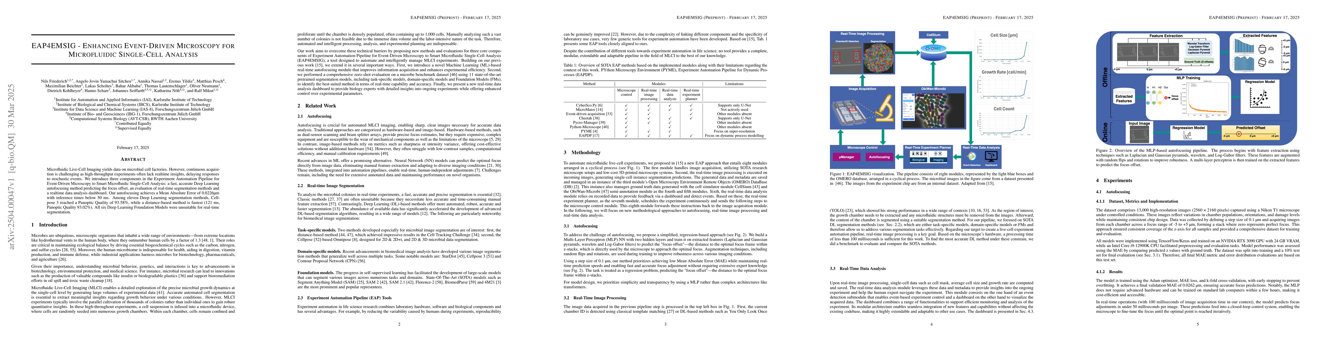
accurate Deep Learning autofocusing method predicting the focus offset, an evaluation of real-time segmentation methods and a realtime data analysis dashboard. Our autofocusing achieves a Mean Absolute Error of 0.0226\textmu m with inference times below 50~ms. Among eleven Deep Learning segmentation methods, Cellpose~3 reached a Panoptic Quality of 93.58\%, while a distance-based method is fastest (121~ms, Panoptic Quality 93.02\%). All six Deep Learning Foundation Models were unsuitable for real-time segmentation.

AI Key Findings

Generated Jun 10, 2025

Methodology

The research introduces an Experiment Automation Pipeline for Event-Driven Microscopy in Smart Microfluidic Single-Cell Analysis, incorporating a fast Deep Learning autofocusing method, an evaluation of real-time segmentation methods, and a realtime data analysis dashboard.

Key Results

  • The proposed Deep Learning autofocusing method achieves a Mean Absolute Error of 0.0226µm with inference times below 50ms.
  • Cellpose~3 among eleven Deep Learning segmentation methods reached a Panoptic Quality of 93.58%.
  • A distance-based segmentation method is fastest (121ms) with a Panoptic Quality of 93.02%.

Significance

This research is important for enhancing microfluidic live-cell imaging in high-throughput experiments, providing real-time insights into stochastic events, and facilitating quicker responses in microbial cell factory studies.

Technical Contribution

The paper presents a novel autofocusing method and a comprehensive evaluation of real-time segmentation techniques for microfluidic single-cell analysis, along with a realtime data analysis dashboard.

Novelty

The work distinguishes itself by combining an accurate, fast Deep Learning autofocusing method with a thorough assessment of real-time segmentation approaches, providing a complete solution for enhancing event-driven microscopy in microfluidic single-cell analysis.

Limitations

  • All six Deep Learning Foundation Models were found unsuitable for real-time segmentation.
  • The study did not explore the impact of varying cell densities or microfluidic channel geometries on segmentation performance.

Future Work

  • Investigate the performance of the proposed methods under varying cell densities and microfluidic channel geometries.
  • Explore the integration of the autofocusing and segmentation methods with other microfluidic platforms.

Paper Details

PDF Preview

Download PDF

Key Terms

No key terms available for this paper.

No citations found for this paper.

Comments (0)

This website uses cookies to ensure you get the best experience. Learn more