Publication
Metrics
AI Quick Summary
The ECG-Image-Database is a comprehensive collection of synthetically generated and physically altered 12-lead ECG images with real-world artifacts, designed to enhance machine learning models for computerized ECG digitization and analysis. The dataset aids in developing robust algorithms to convert ECG images into time-series data, supporting efforts in ECG digitization and annotation.
Paper Preview
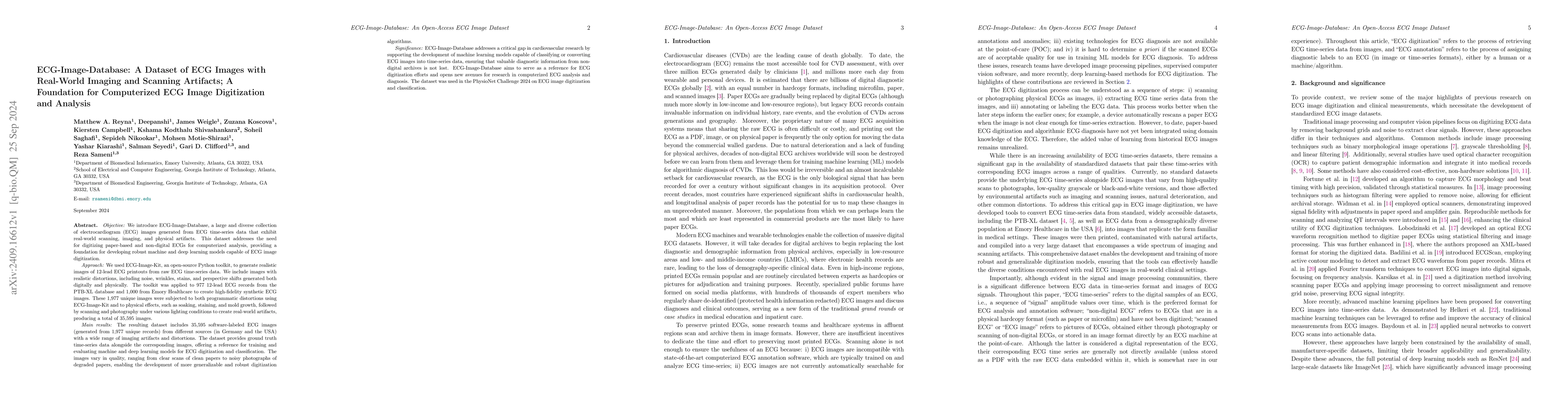
Abstract
We introduce the ECG-Image-Database, a large and diverse collection of electrocardiogram (ECG) images generated from ECG time-series data, with real-world scanning, imaging, and physical artifacts. We used ECG-Image-Kit, an open-source Python toolkit, to generate realistic images of 12-lead ECG printouts from raw ECG time-series. The images include realistic distortions such as noise, wrinkles, stains, and perspective shifts, generated both digitally and physically. The toolkit was applied to 977 12-lead ECG records from the PTB-XL database and 1,000 from Emory Healthcare to create high-fidelity synthetic ECG images. These unique images were subjected to both programmatic distortions using ECG-Image-Kit and physical effects like soaking, staining, and mold growth, followed by scanning and photography under various lighting conditions to create real-world artifacts. The resulting dataset includes 35,595 software-labeled ECG images with a wide range of imaging artifacts and distortions. The dataset provides ground truth time-series data alongside the images, offering a reference for developing machine and deep learning models for ECG digitization and classification. The images vary in quality, from clear scans of clean papers to noisy photographs of degraded papers, enabling the development of more generalizable digitization algorithms. ECG-Image-Database addresses a critical need for digitizing paper-based and non-digital ECGs for computerized analysis, providing a foundation for developing robust machine and deep learning models capable of converting ECG images into time-series. The dataset aims to serve as a reference for ECG digitization and computerized annotation efforts. ECG-Image-Database was used in the PhysioNet Challenge 2024 on ECG image digitization and classification.
AI Key Findings
Get AI-generated insights about this paper's methodology, results, significance, and more — seven facets brought into focus.
Impact
Paper Details
Authors
PDF Preview
Citation Network
Current paper (gray), citations (green), references (blue)
Display is limited for performance on very large graphs.
Discussion 0