Edge-Based Self-Supervision for Semi-Supervised Few-Shot Microscopy Image Cell Segmentation
Publication
Metrics
AI Quick Summary
This paper proposes a semi-supervised learning approach for few-shot microscopy image cell segmentation that leverages edge-based self-supervision to reduce the need for large-scale labelled datasets. The method achieves comparable performance with only 10% of the original training set annotated, demonstrating significant efficiency gains.
Paper Preview
Abstract
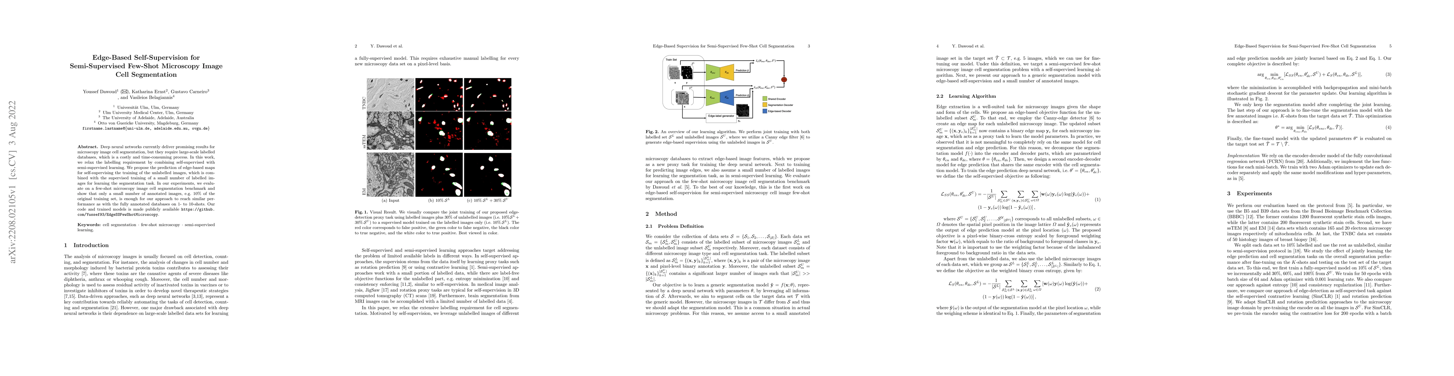
Deep neural networks currently deliver promising results for microscopy image cell segmentation, but they require large-scale labelled databases, which is a costly and time-consuming process. In this work, we relax the labelling requirement by combining self-supervised with semi-supervised learning. We propose the prediction of edge-based maps for self-supervising the training of the unlabelled images, which is combined with the supervised training of a small number of labelled images for learning the segmentation task. In our experiments, we evaluate on a few-shot microscopy image cell segmentation benchmark and show that only a small number of annotated images, e.g. 10% of the original training set, is enough for our approach to reach similar performance as with the fully annotated databases on 1- to 10-shots. Our code and trained models is made publicly available
AI Key Findings
Get AI-generated insights about this paper's methodology, results, significance, and more — seven facets brought into focus.
Impact
Paper Details
Authors
PDF Preview
Key Terms
Citation Network
Current paper (gray), citations (green), references (blue)
Display is limited for performance on very large graphs.
Discussion 0