Effect of adhesion geometry and rigidity on cellular force distributions
Publication
Metrics
AI Quick Summary
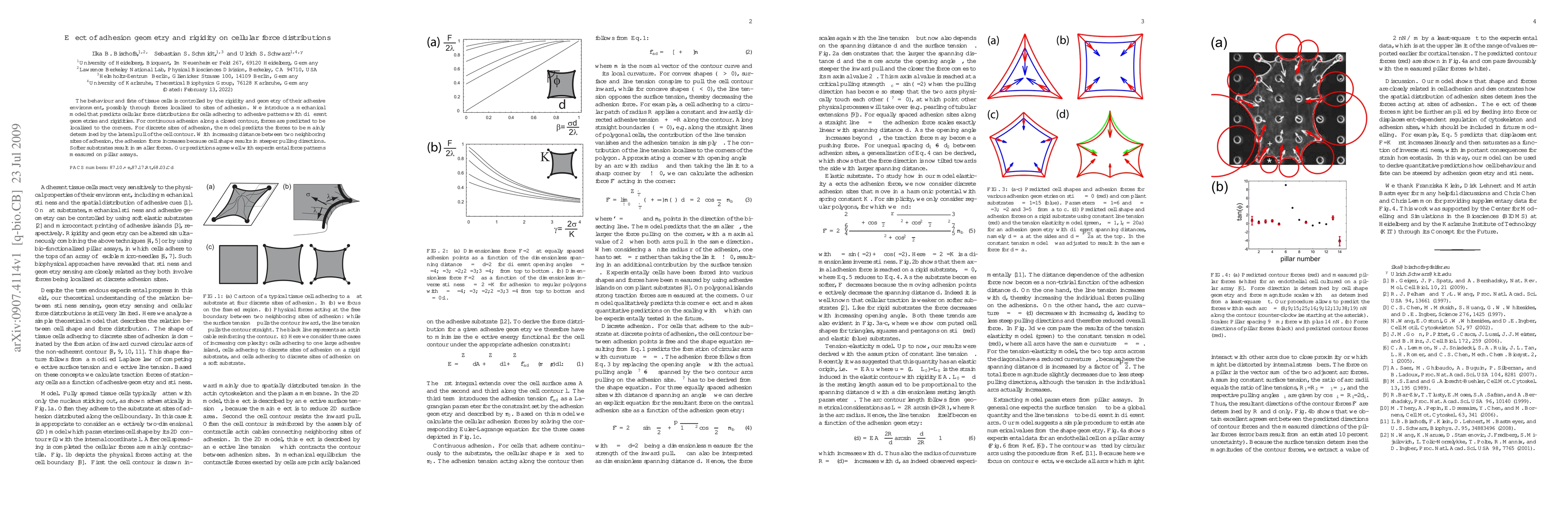
This research introduces a model predicting cellular force distributions based on adhesion geometry and substrate rigidity. The model suggests force concentration at corners for continuous adhesion and at discrete adhesion sites, with forces increasing with greater adhesion site separation. Soft substrates yield smaller forces, aligning with experimental pillar assay results.
Paper Preview
Abstract
The behaviour and fate of tissue cells is controlled by the rigidity and geometry of their adhesive environment, possibly through forces localized to sites of adhesion. We introduce a mechanical model that predicts cellular force distributions for cells adhering to adhesive patterns with different geometries and rigidities. For continuous adhesion along a closed contour, forces are predicted to be localized to the corners. For discrete sites of adhesion, the model predicts the forces to be mainly determined by the lateral pull of the cell contour. With increasing distance between two neighboring sites of adhesion, the adhesion force increases because cell shape results in steeper pulling directions. Softer substrates result in smaller forces. Our predictions agree well with experimental force patterns measured on pillar assays.
AI Key Findings
Get AI-generated insights about this paper's methodology, results, significance, and more — seven facets brought into focus.
Impact
Paper Details
PDF Preview
Key Terms
Citation Network
Current paper (gray), citations (green), references (blue)
Display is limited for performance on very large graphs.
Related Papers
No references found for this paper.
Discussion 0