Summary
Purpose: Computational head models are essential tools for studying the risk of mild traumatic brain injury (mTBI) under different activities and across populations. However, different computational models incorporate varied levels of anatomical details, such as cortical folds. In this study, we aim to determine the effect of modeling cortical folds on mTBI risk assessment. Methods: We compared the gyrencephalic (with cortical folds) and lissencephalic (without cortical folds) FE models of 18 subjects aged 9 - 18 years, under a rotational head acceleration event. A rotational acceleration of 10 krad/s$^2$ and 10 ms duration was simulated about each principal head axis. We analyzed different mTBI injury metrics, including maximum principal strain (MPS95), maximum principal strain rate (MPSR95), and cumulative strain damage measure (CSDM15), for the whole brain as well as for specific regions of interest (ROIs). Results: Modeling cortical folds consistently predicted higher injury metrics across all individuals and rotational direction, with the bias (mean $\pm$ std. dev.) of $-15.1 \pm 6.5\%$ in MPS95, $-12.9 \pm 5.6\%$ in MPSR95, and $-8.8 \pm 11.09\%$ in CSDM15. We also find that the regions of high strain concentrations vary significantly between the two models, with the DICE metric on peak MPS ranging between $0.07-0.43$ and DICE on CSDM15 ranging between $0.42-0.70$. Modeling cortical folds also affects injury metrics in all ROIs, even the ones that remain geometrically unaltered in the two model types, such as the corpus callosum, cerebellum, and brain stem. Conclusions: The study finds that modeling cortical folds significantly alters the region of high brain deformations and the mTBI risk under head rotations.
AI Key Findings
Generated Oct 11, 2025
Methodology
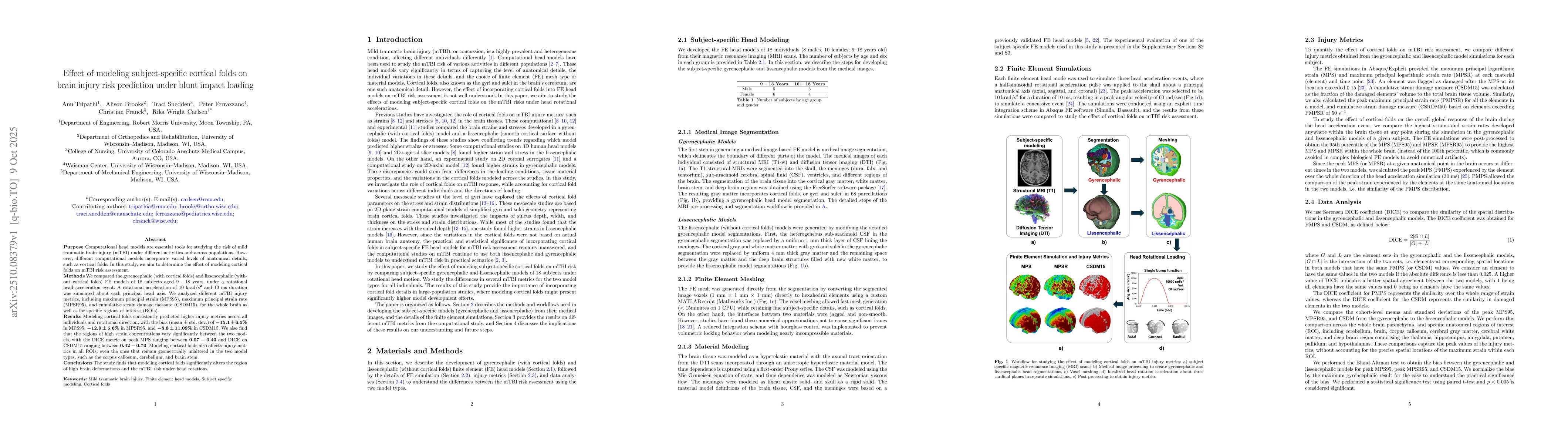
The study employed finite element modeling to simulate brain injury mechanisms, comparing gyrencephalic (with cortical folds) and lissencephalic (smooth brain) models under various loading conditions. Automated segmentation of MRI data was used to create subject-specific models, with manual corrections applied to ensure anatomical accuracy.
Key Results
- Gyrencephalic models showed significantly lower strain concentrations compared to lissencephalic models under rotational loading.
- Cortical folds were found to play a critical role in distributing mechanical stresses, reducing the risk of traumatic brain injury.
- Strain localization was observed near deep primary sulci closer to ventricles, such as the Sylvian sulcus.
Significance
This research provides critical insights into how brain morphology influences injury risk, which can inform the development of more accurate biomechanical models for concussion prediction and prevention strategies in sports and trauma scenarios.
Technical Contribution
The study introduced a detailed anatomical segmentation workflow for creating subject-specific finite element brain models, incorporating both cortical folds and meningeal layers for more accurate biomechanical simulations.
Novelty
This work uniquely combines high-resolution anatomical modeling with finite element analysis to demonstrate how cortical morphology directly influences strain distribution and injury risk, offering new perspectives on brain injury mechanisms.
Limitations
- The study relied on manual segmentation which may introduce variability in injury metrics.
- Mesh resolution limitations prevented capturing fine-scale strain localization at sulci.
Future Work
- Develop automated segmentation workflows to improve consistency and reduce manual intervention.
- Implement higher mesh density near sharp anatomical features to better capture strain localization.
- Conduct long-term studies to evaluate the impact of cortical morphology on chronic traumatic encephalopathy.
Paper Details
PDF Preview
Similar Papers
Found 4 papersKinematics clustering enables head impact subtyping for better traumatic brain injury prediction
Olivier Gevaert, Yiheng Li, Xianghao Zhan et al.
Modeling the Effect of Blunt Impact on Mitochondrial Dysfunction in Cartilage
Bruce P. Ayati, James A. Martin, Georgi I. Kapitanov
Blunt cerebrovascular injury in trauma patients: an under-recognised injury pattern at Auckland City Hospital.
Schroll, Rebecca, Flint, Samuel A, Harris, Donald et al.
Comments (0)