The study irradiated graphene oxide (GO) samples with electron beams at energies of 10, 20, and 30 keV using a scanning electron microscope (SEM), varying exposure times from 15 minutes to 1 hour. Raman D- and G-band intensities were analyzed to assess structural modifications.
Publication
Metrics
AI Quick Summary
This study investigates the impact of electron-beam irradiation from a scanning electron microscope on graphene oxide films, revealing that a 30 keV beam for 30 minutes can induce amorphous carbon formation, while lower energies (10 keV and 20 keV) for similar durations do not significantly alter the structural integrity of the graphene oxide.
Quick Answers
What is "Effects of Electron-Beam Irradiation on Graphene Oxide" about?
This study investigates the impact of electron-beam irradiation from a scanning electron microscope on graphene oxide films, revealing that a 30 keV beam for 30 minutes can induce amorphous carbon formation, while lower energies (10 keV and 20 keV) for similar durations do not significantly alter the structural integrity of the graphene oxide.
What methodology did the authors use?
The study irradiated graphene oxide (GO) samples with electron beams at energies of 10, 20, and 30 keV using a scanning electron microscope (SEM), varying exposure times from 15 minutes to 1 hour. Raman D- and G-band intensities were analyzed to assess structural modifications. More in Methodology →
What are the key results?
30 keV electron beam irradiation for 30 minutes causes formation of amorphous carbon in GO. — 10 keV and 20 keV beams for 30 minutes do not significantly affect GO samples. More in Key Results →
Why is this work significant?
This research is crucial for understanding the impact of SEM imaging on GO properties, which is essential for accurate characterization and potential applications in materials science. More in Significance →
What are the main limitations?
Limited irradiation time intervals for long-term effects analysis. — Lack of comprehensive understanding of amorphous carbon formation mechanisms under prolonged irradiation. More in Limitations →
Abstract
Graphene oxide (GO) is a nanofilm composed of graphene with various oxygen functional groups attached. GO is of interest due to its unique mechanical-enhancement properties, its tunable electronic properties, and its potential use in the wide-scale production of graphene. Scanning electron microscopes (SEMs) are frequently used to characterize and study GO films. The purpose of this project was to study the effects of SEM-imaging on GO films. Using an SEM, we irradiated GO samples at electron beam-energies of 10, 20, and 30 keV (at a constant emission current of ~40 micro-amps) for times ranging from 15 minutes to one hour. Raman D- and G-band intensities were used to examine structural modifications/damage to GO samples as a function of beam energy and exposure time. The results suggest that imaging with a 30 keV electron beam for 30 minutes may lead to the formation of amorphous carbon, while imaging with 10 keV or 20 keV beams for 30 minutes does not have a significant effect on GO samples.
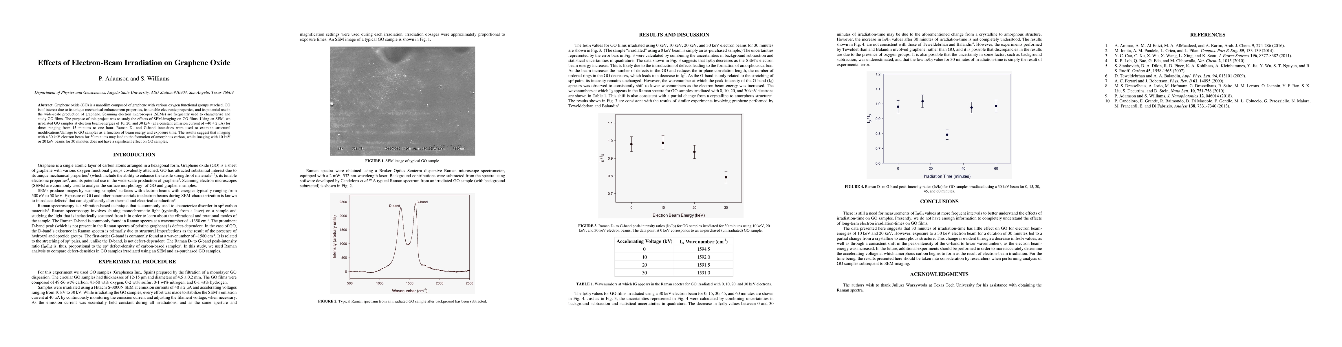
Key Findings, in focus
Seven facets of this paper, analysed and brought into focus by AI.
This research is crucial for understanding the impact of SEM imaging on GO properties, which is essential for accurate characterization and potential applications in materials science.
- 30 keV electron beam irradiation for 30 minutes causes formation of amorphous carbon in GO.
- 10 keV and 20 keV beams for 30 minutes do not significantly affect GO samples.
- ID/IG ratio decreases with increasing electron beam energy, indicating a crystalline to amorphous structure transition.
This research is crucial for understanding the impact of SEM imaging on GO properties, which is essential for accurate characterization and potential applications in materials science.
Detailed analysis of electron-beam irradiation effects on graphene oxide, providing insights into structural changes and their correlation with beam energy and exposure time.
This work distinguishes itself by focusing on the impact of SEM imaging on GO, offering specific data on electron-beam energy and exposure time dependencies, which is valuable for optimizing SEM use in GO characterization.
- Limited irradiation time intervals for long-term effects analysis.
- Lack of comprehensive understanding of amorphous carbon formation mechanisms under prolonged irradiation.
- Investigate effects of longer irradiation times to establish a clearer timeline for structural changes.
- Explore the accelerating voltage threshold for amorphous carbon formation in GO.
Impact
Paper Details
PDF Preview
Key Terms
Citation Network
Current paper (gray), citations (green), references (blue)
Display is limited for performance on very large graphs.
Discussion 0